

Search results: TNFRSF13C
Product group Assays
Rat TNFRSF13C/BAFFR ELISA KitRTFI00590
Assay Sample TypeSerum, Plasma
ReactivityRat
- SizePrice
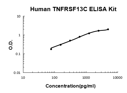
Product group Assays
Assay Sample TypeSerum, Plasma
ReactivityHuman
- SizePrice
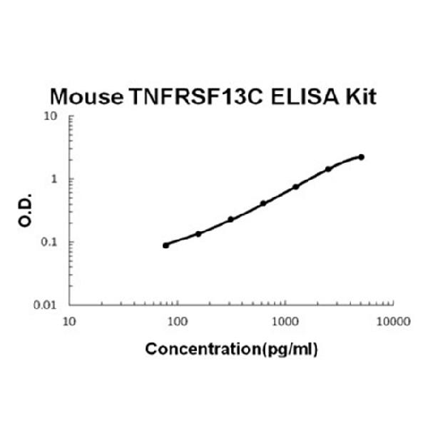
Product group Assays
Assay Sample TypeSerum, Plasma
ReactivityMouse
- SizePrice
Product group Antibodies
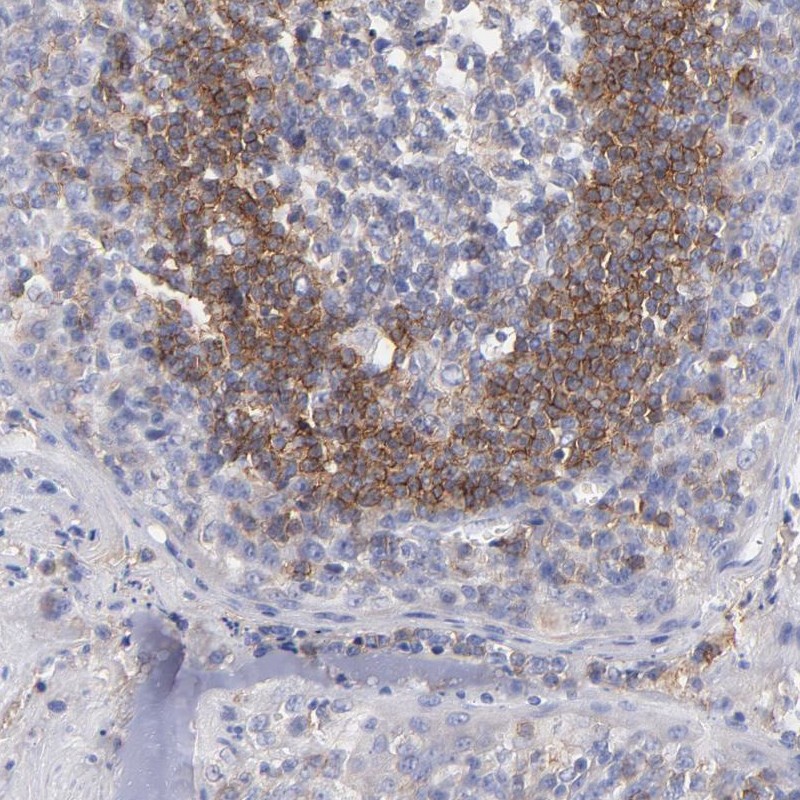
TNFRSF13C antibodyORB607235
ApplicationsImmunoHistoChemistry, ImmunoHistoChemistry Paraffin
ReactivityHuman
TargetTNFRSF13C
- SizePrice
Product group Antibodies
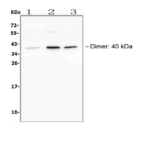
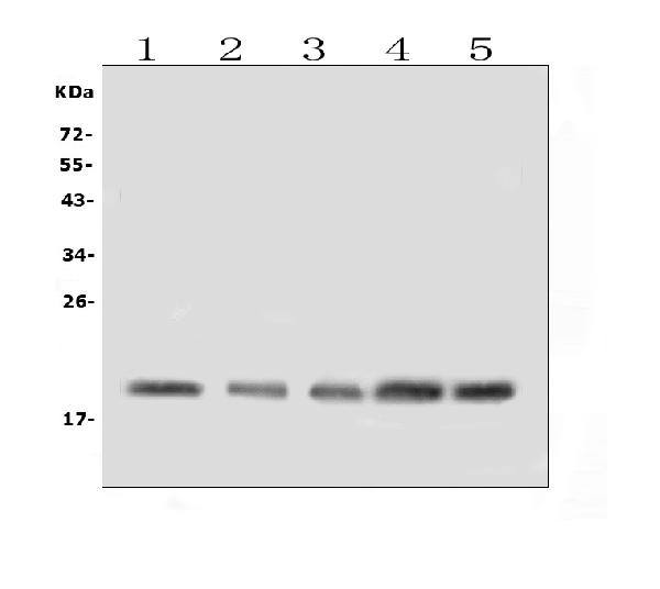
Anti-TNFRSF13CHPA003246
ApplicationsWestern Blot, ImmunoHistoChemistry
ReactivityHuman
TargetTNFRSF13C
- SizePrice
Product group Assays
- SizePrice
Product group Antibodies
TNFRSF13C Polyclonal AntibodyRD79178A
ApplicationsELISA, ImmunoHistoChemistry
ReactivityHuman, Mouse
TargetTNFRSF13C
- SizePrice
Product group Assays
- SizePrice