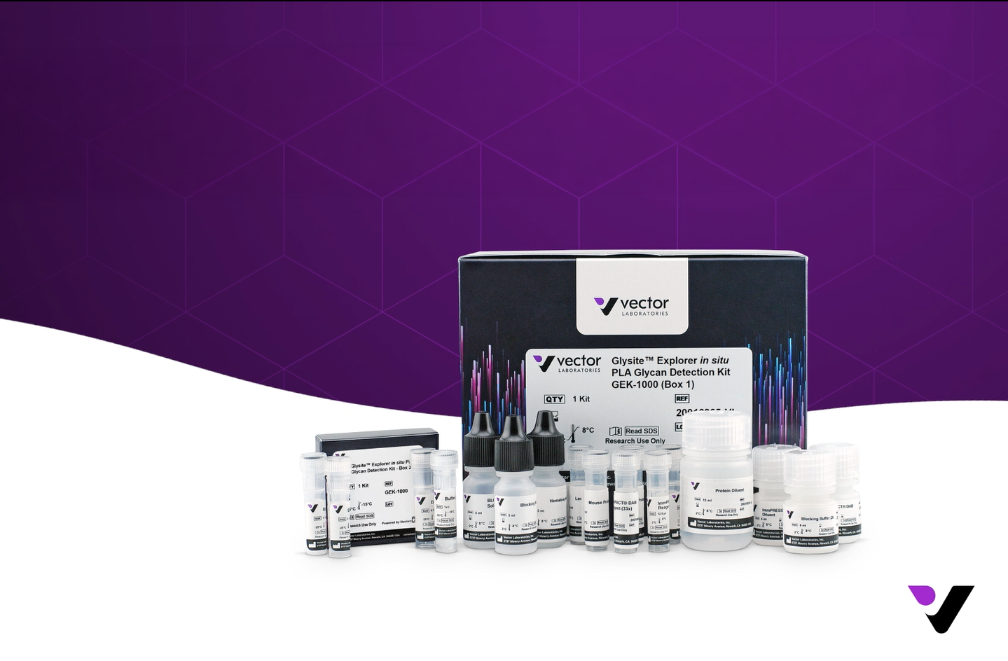
Vector Laboratories‘ Glysite™ Explorer in situ PLA Glycan Detection Kit is now available through Bio-Connect. This first-of-its-kind tool transforms the way researchers approach spatial glycosylation.
The field of spatial biology just gained a powerful new dimension. Vector Laboratories has launched the Glysite™ Explorer in situ PLA Glycan Detection Kit, a first-of-its-kind solution for visualizing protein glycosylation directly in tissue and cell samples. Bio-Connect is proud to announce that this innovative kit is now available through our portfolio.
In this article, you will find what makes this kit unique, how it works, what it detects, which sample types it supports, and why it matters for your research.
Protein glycosylation is one of the most prevalent and functionally important post-translational modifications in biology. It influences protein folding, stability, cellular signaling, and immune recognition, and it plays a central role in health and disease, from cancer biology to infectious diseases. Despite its significance, studying glycosylation in a spatial, tissue-level context has historically been challenging. The Glysite™ Explorer in situ PLA Glycan Detection Kit from Vector Laboratories changes that.
This kit is the first of its kind to combine lectin-based glycan detection with in situ Proximity Ligation Assay (isPLA) technology, enabling researchers to visualize where specific glycans occur in proximity to a protein of interest, directly within FFPE tissue sections, FFPE cell pellets, and fixed cells.
The Glysite™ Explorer in situ PLA Glycan Detection Kit (catalog number GEK-1000) integrates two proven technologies: Vector Laboratories’ curated panel of Glysite™ Explorer Lectins and Navinci’s proprietary isPLA platform.
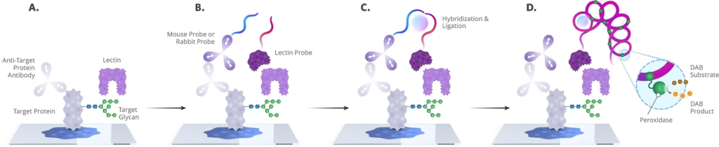
The workflow is straight forward:
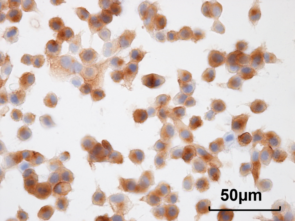
The result is a highly sensitive, spatially resolved signal that reveals the glycosylation status of a target protein in its native tissue environment.
The kit is designed to work with a flexible selection of Glysite™ Explorer Lectins, covering eight major glycan categories. Researchers can choose one or more lectins (up to ten) to address their specific biological question:
| Lectin | Glycan Specificity |
| WGA Lectin | Terminal GlcNAc-containing glycans |
| LCA Lectin | α1-6 fucose |
| PHA-L Lectin | β1-6 branched N-glycans (tri- and tetraantennary) |
| ECL Lectin | Terminal type 2 LacNAc / LacdiNAc |
| Jacalin Lectin | Core 1 and Core 3 O-glycans |
| GNL Lectin | Terminal α1-3 or α1-6 mannose |
| MAL II Lectin | α2-3 sialylated glycans |
| SNA Lectin | α2-6 sialylated LacNAc / LacdiNAc |
| WFA Lectin | Terminal GalNAc and multiantennary LacNAc |
| AAL Lectin | α-fucose |
Each lectin is available separately (50x concentrated, sufficient for 50 reactions) and is simply diluted with Protein Diluent prior to use.
One of the key advantages of the Glysite™ Explorer Kit is how naturally it fits into existing laboratory workflows. It is designed to plug directly into standard IHC and isPLA protocols without requiring specialized equipment, additional training, or complex setup. The optimized protocol can be completed within a single day, approximately 8 hours when using a 30-minute primary antibody incubation.
The kit is compatible with multiple peroxidase substrates, including the included ImmPACT® DAB, as well as ImmPACT® NovaRed, ImmPACT® VIP, and ImmPACT® SG. Slides can be mounted with both aqueous and non-aqueous mounting media, such as VectaMount® PT or VectaMount® AQ.
The Glysite™ Explorer in situ PLA Glycan Detection Kit has been validated on the following sample types:
This broad compatibility makes it possible to leverage existing archived sample collections for retrospective glycosylation studies, as well as freshly processed specimens. Optimal section thickness is 4 to 6 µm, and the kit provides 5 mL of working solution, sufficient to stain 50 to 100 specimens.
The kit is validated on human specimens, and other species are expected to be compatible provided that appropriate primary antibody species are selected to avoid endogenous IgG interference.
The Glysite™ Explorer Kit is already making an impact in cutting-edge cancer research. Dr. Steven Barthel and Dr. Tobias Schatton, who co-direct the Program of Glyco-Immunology and Oncology (PGIO) at Brigham and Women’s Hospital, Harvard Medical School, are using the kit to explore glycan proximity to immune checkpoint proteins in melanoma cells and diverse immune cell subsets within the tumor microenvironment. Their work is contributing to a deeper understanding of how glycosylation influences cancer progression, in research that would not have been feasible without this technology.
The Glysite™ Explorer in situ PLA Glycan Detection Kit is ideal for researchers working in:
The Glysite™ Explorer in situ PLA Glycan Detection Kit (GEK-1000) is now available through Bio-Connect. The base kit includes all core detection reagents, and Individual Glysite™ Explorer Lectins are available separately to complement your assay.
Is this the kit for your research? Order it directly via our webshop or reach out to your personal account manager. We are happy to help you select the right Glysite™ Explorer Lectins for your specific research application.
Antibodies | Glycobiology | Immunodetection Reagents | Specialty Chemicals | Mounting Medium | Staining
We gladly support you by keeping you updated on our latest products and developments