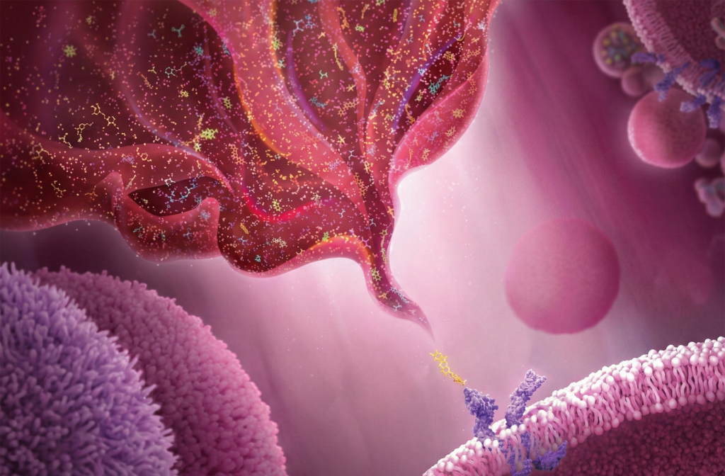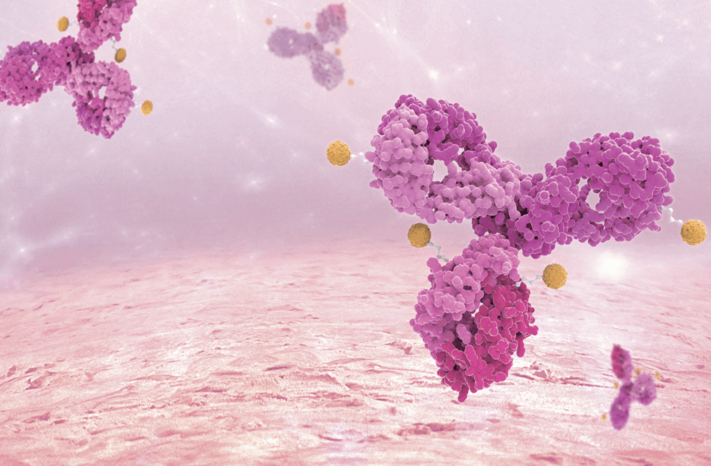
In immune cell culture, high-quality cytokines from MedChem Express unlock the potential of cells, guiding their fate with precision and enabling more efficient, reliable experimental outcomes.
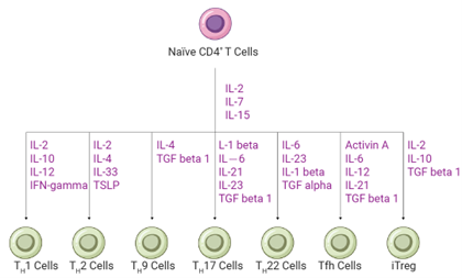
T cells are central to adaptive immunity, and their function and differentiation are determined by characteristic surface markers, with the most important being the T cell receptor (TCR) and differentiation markers (CD molecules). Based on TCR types, T cells can be divided into two main subgroups: αβ T cells and γδ T cells. Dominant αβ T cells recognize antigens presented by the cell’s own MHC (Major Histocompatibility Complex) molecules and differentiate into two subtypes: CD4+ T cells, which function as “helper” T cells, regulating global immune responses by coordinating antigen-presenting cells (APCs), and CD8+ T cells, which act as “cytotoxic” T cells, directly recognizing and killing abnormal target cells. Unlike conventional αβ T cells, γδ T cells are a special subgroup of T cells that occupy only a small portion of peripheral blood (0.5%-5%) but have unique functions. They can recognize a variety of antigens without MHC presentation, combining the rapid response of innate immunity with the memory function of adaptive immunity. This characteristic gives them enormous potential in anti-tumor immunity[1][2].
Whether it is conventional αβ T cells or special γδ T cells, their functional performance relies on precise regulatory mechanisms. High-quality cytokines from MedChem Express provide reliable tools to modulate these pathways in vitro. For example, the differentiation of naïve CD4+ T cells after activation is highly dependent on cytokine signals in the local microenvironment. A specific combination of cytokines activates different transcription factors (such as T-bet, GATA3, RORγt, FoxP3, Bcl-6, etc.), driving distinct differentiation programs[1].
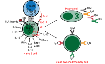
B cells express B cell receptors (BCRs), which can specifically bind to antigens and produce antibodies. In vitro, B cells can undergo class switching and differentiation in response to various cytokine stimuli, ultimately becoming antibody-secreting cells that produce antibodies and secrete regulatory cytokines[4].
This process begins with the critical co-stimulatory signal provided by CD40L, which mimics the helper role of T cells and is fundamental for B cell activation and proliferation. IL-4 is the core factor driving B cell proliferation and inducing antibody class switching (especially to IgE and IgG1). IL-21 strongly promotes B cell differentiation into plasma cells and regulates antibody secretion. The survival and homeostasis of B cells are governed by BAFF and APRIL, which are key to maintaining mature B cell and long-lived plasma cell survival. Additionally, other factors play important roles in specific contexts: IFN-γ induces class switching to IgG2a (mouse) / IgG1 (human); IL-6 and IL-10 support plasma cell differentiation and survival; and IL-2, IL-7, and IL-15 primarily provide proliferative support signals[5][6].
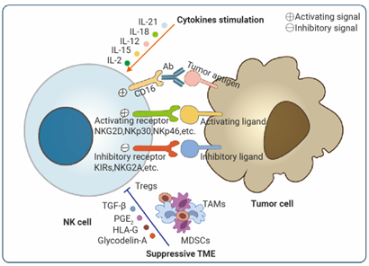
Natural Killer (NK) cells are different from other lymphocytes (such as B cells, T cells, and NKT cells) in that they do not express antigen-specific receptors like BCRs or TCRs. Instead, NK cells function in an antigen-independent manner, possessing intrinsic cytotoxic potential and cytokine production capacity[7].
The in vitro expansion, activation, and functional maintenance of NK cells depend on a finely tuned cytokine network. IL-15 is the most critical factor, driving NK cell development, survival, proliferation, and enhancing cytotoxicity. To generate more functional NK cells, IL-15 is often combined with IL-12 and IL-18, which together strongly induce IFN-γ secretion and significantly enhance their anti-tumor and anti-viral capabilities[8]. IL-21 can regulate the differentiation state of NK cells, enhancing their antibody-dependent cellular cytotoxicity (ADCC) and is commonly used in CAR-NK cell preparations. Furthermore, type I interferons (IFN-α/β) can directly activate NK cells. When inducing NK cells from hematopoietic stem cells or progenitor cells, FLT3L and IL-3 are required to expand and maintain early precursor cell pools. It is important to note that TGF-β and IL-10 are potent inhibitors of NK cell function and are often found in tumor microenvironments. These factors need to be neutralized in vitro to optimize NK cell function[9][10][11].
Monocytes are the “reserves” in the blood, which migrate to tissues and differentiate into “resident troops”, macrophages (M0) or dendritic cells. They are central to innate immunity, performing three main functions: phagocytosis of pathogens, antigen presentation to activate adaptive immunity, and cytokine secretion to direct global immune responses.
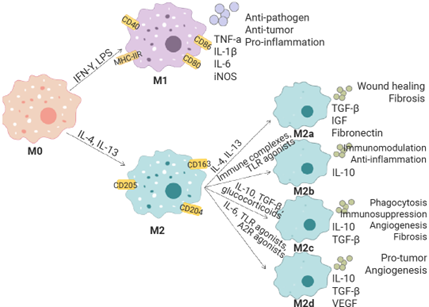
In vitro, various cytokines are needed to maintain monocytes, activate their differentiation, and expand them into macrophages and their subtypes. For example, human macrophages are derived from purified CD14+ monocytes and are differentiated into M0 macrophages using M-CSF. Specific polarization factors further guide them into M1 and M2 macrophages. LPS combined with IFN-γ drives polarization to the M1 phenotype (pro-inflammatory), while IL-4 and IL-13 induce polarization to the M2 phenotype (anti-inflammatory). Each macrophage polarization state shows unique cytokine profiles and surface markers, which are typically verified by ELISA or flow cytometry (for example, M1 macrophages upregulate CD86, MHC II, etc.). This allows characterization of macrophage populations under different tissue inflammation conditions, which helps to understand disease pathogenesis[13][14][15].
Dendritic cells (DCs) are a group of hematopoietic cells that serve as a bridge between the innate and adaptive immune systems. Their most important function is processing and presenting antigens to T cells, making them the most effective antigen-presenting cells currently known, and an important target for cancer immunotherapy[16]. Various methods for culturing and expanding DCs from different sources (e.g., bone marrow, peripheral blood monocytes, induced pluripotent stem cells) have been described in the literature. Regardless of the source, the in vitro culture, activation, and maturation of DCs require a cooperative action of various cytokines. For example, FLT-3 Ligand, SCF, and GM-CSF are decisive in the development of blood-derived DC subgroups (classical and plasmacytoid).
| Cell Source | Core Cytokines Required for Induction | Scheme Characteristics and Advantages |
| Peripheral Blood Monocytes | Immature DCs:GM-CSF + IL-4 Mature DCs: TNF-α + IL-1β + IL-6 + PGE2 Tolerogenic DCs: IL-10 + TGF-β | Classic, widely used protocol. Short cycle (7–9 days), high yield, mature technique. |
| CD34+ Hematopoietic Stem Cells (HSC) | FLT-3 Ligand (core) ± GM-CSF, SCF, TNF-α | Can generate multiple subgroups, more representative of in vivo DC heterogeneity. Longer cycle (14–21 days), complex protocol. |
| Direct Isolation | No induction, direct isolation | Direct sorting from blood or tissue. Highest purity, closest to natural state, but low yield. |
| Pluripotent Stem Cells (iPSC/ESC) | Various factors added in stages (e.g., BMP-4, VEGF, SCF, FLT-3 Ligand, GM-CSF) | Unlimited expansion, stable source, gene-editable. Complex differentiation protocol, long cycle (28+ days), high cost. |
Granulocytes are a type of white blood cell containing granules in their cytoplasm, and they are classified into neutrophils, eosinophils, and basophils based on their staining characteristics[22]. Culturing and differentiating granulocytes, particularly primary neutrophils, in vitro is highly challenging due to their terminal differentiation status, short lifespan, and extremely low proliferation capacity in vitro. Currently, mature granulocytes are primarily obtained by directly isolating them from peripheral blood or bone marrow, or by directed differentiation from CD34+ hematopoietic stem cells. IL-3, an effective hematopoietic growth factor, participates in the differentiation of bone marrow progenitor cells and the activation of basophils. IL-5 can activate eosinophils and, together with IL-3 and GM-CSF, promotes their development and maturation[23][24].
Maintain Cell Viability: Recombinant cytokines help sustain immune cell survival while simultaneously activating and differentiating the cells to reach the desired phenotype.
Optimize Cell Function: These cytokines provide the necessary signals to enhance immune cell activation, proliferation, differentiation, and survival, thereby improving their adaptability and functionality in the in vitro environment.
Standardize Culture Conditions: Recombinant cytokines possess high purity, biological activity, low endotoxin levels, and batch-to-batch consistency, ensuring a standardized culture environment. This standardization minimizes variability in experimental results, enhancing reproducibility and reliability.
MedChem Express provides a comprehensive range of high-quality cytokines for immune cell culture, along with culture media, sorting magnetic beads, antibodies, agonists, and other related products to meet your experimental needs and support your research.
| IL-2 | High-dose expansion of effector T cells (CAR-T); low-dose maintenance of Treg function. |
| TGF-β1 | Induces Treg or Th17 differentiation at low concentrations; inhibits immune response and promotes fibrosis at high concentrations; induces B cell switching to IgA class. |
| IFN-γ | Classical activation of M1 macrophages; enhances antigen presentation; promotes Th1 response. |
| GM-CSF | Induces monocyte differentiation into macrophages (predominantly M1) or dendritic cells (DC); supports eosinophil generation in combination with IL-5. |
| TNF-α | Induces DC maturation, upregulates co-stimulatory molecules (CD80/86) and MHC-II expression; pro-inflammatory. |
| CD40L | Provides the essential second signal for B cell activation, driving proliferation, germinal center formation, and class switching; promotes DC maturation. |
| IL-1β | Co-operates in inducing Th17 differentiation; activates inflammasomes. |
MedChem Express cytokines and related immune cell culture reagents are available through our webshop, where you can easily explore and compare products for your assays. For technical details or experimental advice, please contact our Technical Support team or reach out directly to your account manager.
[1] Dong C. Cytokine Regulation and Function in T Cells. Annu Rev Immunol. 2021 Apr 26;39:51-76.
[2] Silva-Santos B, et al. γδ T cells: pleiotropic immune effectors with therapeutic potential in cancer. Nat Rev Cancer. 2019 Jul;19(7):392-404.
[3] Jiang H, et al. T Cell Subsets in Graft Versus Host Disease and Graft Versus Tumor. Front Immunol. 2021 Oct 5;12:761448.
[4] Cooper MD. The early history of B cells. Nat Rev Immunol. 2015 Mar;15(3):191-7.
[5] Marasco E, et al. B-cell activation with CD40L or CpG measures the function of B-cell subsets and identifies specific defects in immunodeficient patients. Eur J Immunol. 2017 Jan;47(1):131-143.
[6] Moens L, et al. Cytokine-Mediated Regulation of Plasma Cell Generation: IL-21 Takes Center Stage. Front Immunol. 2014 Feb 18;5:65.
[7] Hammer Q, et al. Natural killer cell specificity for viral infections. Nat Immunol. 2018 Aug;19(8):800-808.
[8] Romee R, et al. Cytokine activation induces human memory-like NK cells. Blood. 2012 Dec 6;120(24):4751-60.
[9] Vivier E, et al. Innate or adaptive immunity? The example of natural killer cells. Science. 2011 Jan 7;331(6013):44-9.
[10] Horowitz A, et al. Activation of natural killer cells during microbial infections. Front Immunol. 2012 Jan 3;2:88.
[11] Gaggero S, et al. Cytokines Orchestrating the Natural Killer-Myeloid Cell Crosstalk in the Tumor Microenvironment: Implications for Natural Killer Cell-Based Cancer Immunotherapy. Front Immunol. 2021 Jan 29;11:621225.
[12] Xiao J, et al. Natural Killer Cells: A Promising Kit in the Adoptive Cell Therapy Toolbox. Cancers (Basel). 2022 Nov 17;14(22):5657.
[13] Yunna C, et al. Macrophage M1/M2 polarization. Eur J Pharmacol. 2020 Jun 15;877:173090.
[14] Xia T, et al. Advances in the role of STAT3 in macrophage polarization. Front Immunol. 2023 Apr 4;14:1160719.
[15] Gao J, et al. Shaping Polarization Of Tumor-Associated Macrophages In Cancer Immunotherapy. Front Immunol. 2022 Jun 30;13:888713.
[16] Palucka K, et al. Cancer immunotherapy via dendritic cells. Nat Rev Cancer. 2012 Mar 22;12(4):265-77.
[17] Luo XL, et al. The quest for faithful in vitro models of human dendritic cells types. Mol Immunol. 2020 Jul;123:40-59.
[18] Karsunky H, et al. Flt3 ligand regulates dendritic cell development from Flt3+ lymphoid and myeloid-committed progenitors to Flt3+ dendritic cells in vivo. J Exp Med. 2003 Jul 21;198(2):305-13.
[19] Patente TA, et al. Human Dendritic Cells: Their Heterogeneity and Clinical Application Potential in Cancer Immunotherapy. Front Immunol. 2019 Jan 21;9:3176.
[20] Xiao Z, et al. Dendritic cells instruct T cell anti-tumor immunity and immunotherapy response[J]. The Innovation Medicine, 2025, 3(2): 100128-1-100128-18.
[21] Fucikova J, et al. Induction of Tolerance and Immunity by Dendritic Cells: Mechanisms and Clinical Applications. Front Immunol. 2019 Oct 29;10:2393.
[22] Pascal M, et al. Granulocytes and mast cells in AllergoOncology-Bridging allergy to cancer: An EAACI position paper. Allergy. 2024 Sep;79(9):2319-2345.
[23] Martinez-Moczygemba M, et al. Biology of common beta receptor-signaling cytokines: IL-3, IL-5, and GM-CSF. J Allergy Clin Immunol. 2003 Oct;112(4):653-65; quiz 666.
[24] Tsioumpekou M, et al. The Role of Cytokines in Neutrophil Development, Tissue Homing, Function and Plasticity in Health and Disease. Cells. 2023 Jul 31;12(15):1981.


Inhibitors | Agonists | Recombinant Proteins | Dyes | Assay Kits | Compound Libraries | Drug Screening | PCR | Cell Culture Reagents | Custom Synthesis, Screening, ADC, PROTAC Solutions
We gladly support you by keeping you updated on our latest products and developments