Innovative Tools Driving Progress in Neuroscience Research
Genetically engineered cell lines can serve as excellent model systems in a variety of applications for neuroscience research. BPS Bioscience‘s selection of novel cell lines enables new cellular insights into signaling pathways and drug discovery.
Innovative Tools Driving Progress in Neuroscience Research
Neuroscience is a multidisciplinary field dedicated to studying the nervous system, including the brain, spinal cord, and peripheral nerves. Pharmaceutical companies are developing drugs targeting neurological conditions characterized by cognitive and motor dysfunction due to neuronal or nerve damage, such as Alzheimer’s and Parkinson’s diseases. They also focus on neuropsychiatric conditions, which involve disturbances in behavior and emotional regulation, including anxiety, depression, and addiction.
The terms mental health and mental illness are often used to describe neuropsychiatric disorders. These disorders affect more than 970 million patients globally and are among the leading causes of disability, with an estimated annual economic burden exceeding USD 800 billion. Drug development in neuroscience presents unique challenges, such as the need to bypass the blood brain barrier, and the lack of cellular models. Nonetheless, the global neuroscience market is expanding, driven by an increasing number of biotech companies entering the field and a surge in mergers and acquisitions over the past five years. More than 30,000 scientists are currently engaged in neuroscience-related research worldwide.
Muscarinic receptor cell lines
Muscarinic receptors regulate fundamental physiological functions including heart rate, motor control, cognition, memory, and learning, with their dysfunction resulting in multiple neurological and psychiatric disorders. The evidence shows muscarinic receptors (M1-M5 subtypes) transduce signals through G-protein coupling, with odd-numbered receptors (M1, M3, M5) activating phospholipase C and even-numbered receptors (M2, M4) inhibiting adenylyl cyclase C. Loss of muscarinic receptor function has been implicated in Alzheimer’s disease (AD), Down’s syndrome, and Parkinson’s disease (PD). Muscarinic receptors targeting has resulted in limited but promising therapeutic evidence. Muscarinic allosteric modulators have entered clinical trials for Alzheimer’s disease and schizophrenia, and multiple studies support M1 and M4 receptors as targets for cognitive and behavioral symptoms across psychiatric and neurological disorders. Cobenfy, which targets both M1 and M4 receptors, was the first new schizophrenia treatment to receive approval in more than three decades. In contrast emraclidine, which is specific for the M4 receptor, failed to show efficacy in Phase II trials for schizophrenia.
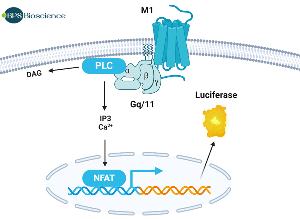
We have generated three cell lines each expressing one of the receptors. The Muscarinic Acetylcholine Receptor (mAChR) M1, M3, and M5/NFAT Luciferase Reporter HEK293 Cell Lines are HEK293 cell lines expressing the full-length human muscarinic acetylcholine receptor M1, M3 or M5, and a firefly luciferase reporter under the control of the NFAT (nuclear factor of activated T cells) response element. These cell lines have been validated for expression of the cogent receptor by flow cytometry, and for their response to agonist Oxotremorine M, showing that luciferase activity increases in proportion to mAChR activation.
Figure 1: Mechanism of luciferase activation in Muscarinic Acetylcholine Receptor (mAChR) M1, M3 or M5 / NFAT Luciferase Reporter HEK293 Cell Lines.
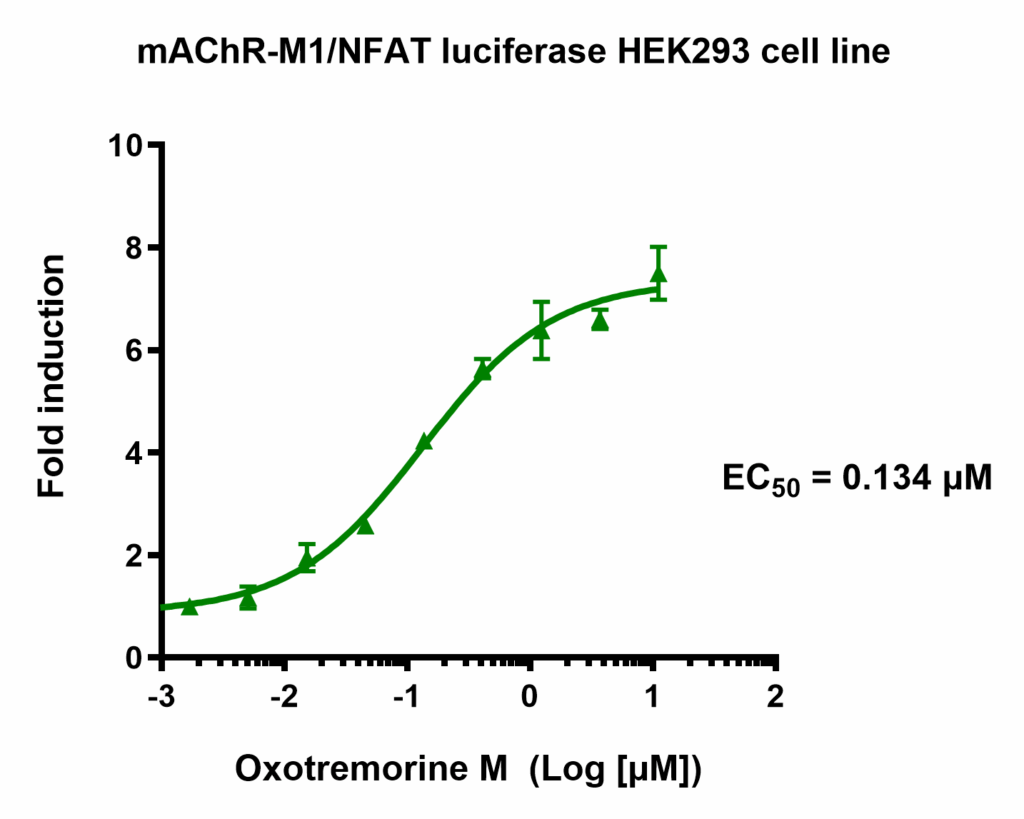
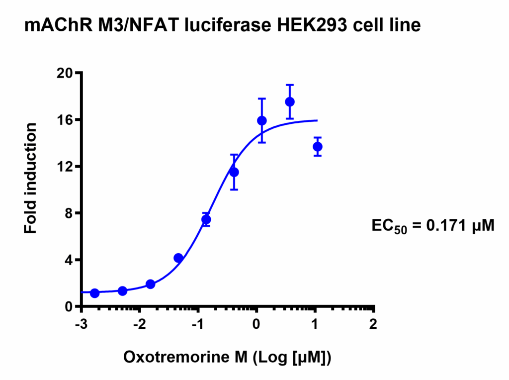
Figure 2: Dose response to Oxotremorine in (mAChR) M1 (left) and M3 (right) NFAT Luciferase Reporter HEK293 Cell Lines.
The Neuro2A cell line is a genetically tractable model widely used in neuroscience for screening and mechanistic studies, which has been instrumental in advancing the understanding of receptor pharmacology, protein trafficking, and the molecular mechanisms underlying genetic neurological disorders.
Although these cells lack certain key neuronal markers, they can undergo dopaminergic differentiation, exhibiting elevated tyrosine hydroxylase expression and increased dopamine production. Furthermore, streptozotocin treatment induces pathological features relevant to Alzheimer’s disease, including tau phosphorylation, amyloid aggregation, and mitochondrial stress, which can be ameliorated by donepezil treatment.
The ability to genetically engineer Neuro2A cells enables the assessment of new drug candidates in cells expressing clinically relevant genetic mutations.
Our Cas9-expressing Neuro2A cells express the endonuclease Cas9 (Streptococcus pyogenes CRISPR associated protein 9) , which is used to introduce the desired modification. Thus, the cells can be transduced or electroporated with sgRNA targeting a gene of interest to quickly generate knock-out cell pools or cell lines.
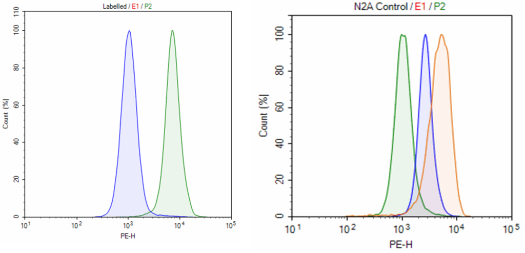
Figure 3: Cas9 expression in Neuro2A cell pool (left #78087) and low expression cell line (right #78137).
iPSC-Derived Cells
Induced pluripotent stem cells (iPSCs) are a powerful tool as they can differentiate into a wide variety of cell types, including neurons. They are used to study disease mechanisms, support drug discovery and development, and advance regenerative medicine applications. Importantly, iPSCs can be generated directly from patient-derived cells, enabling the creation of disease-specific models that provide critical insight into disease progression and facilitate the development of targeted therapeutic strategies.
The Wnt signaling pathway is a well described regulator of organism development and cell fate specification. Cellular pluripotency and differentiation of multiple lineages, including neurons, are modulated by this pathway. In its canonical pathway, cell activation by a Wnt ligand leads to the stabilization and nuclear translocation of β-catenin and subsequent activation of TCF/LEF (T-cell factor/lymphoid enhancing factor) transcription factor. The Wnt signaling pathway is a therapeutic target in the treatment of neuropsychiatric disorders.
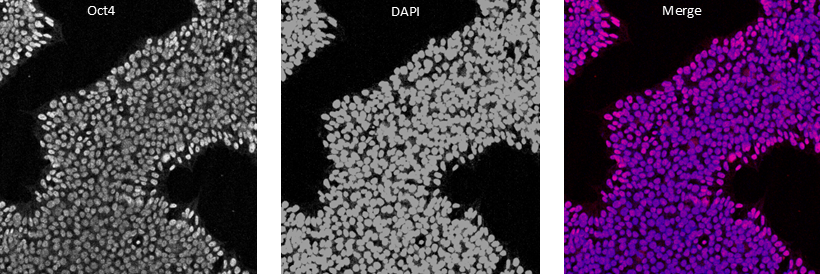
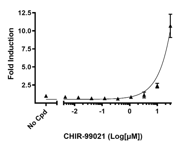
The TCF/LEF StemBright™ Luciferase IPS Cell Pool contain a firefly luciferase reporter under the control of TCF/LEF responsive elements, stably integrated into iPS cells. TCF/LEF transcription factor is downstream of the Wnt signaling pathway. This cell pool is validated for its response to GSK3β inhibitor CHIR-99021, which activates the Wnt signaling pathway.
Figure 4: Immunofluorescence staining of Oct-4 in TCF/LEF StemBright™ Luciferase iPS Cell Pool: iPS Cell marker Oct-4 (left panel), DAPI (middle panel), and merge(purple, right panel).
Figure 5: Dose response of TCF/LEF StemBright™ Luciferase iPS Cell Pool to the GSK3-β inhibitor CHIR-99021.
Resources
Webinar (YouTube)
Reverse engineering the human heart with pluripotent stem cells
Innovative Tools Driving Progress in Neuroscience Research

 Find products for your research