From identifying specific populations of cells, understanding the development of our brain to tracking the progression of neurodegenerative diseases, neural lineage markers has revolutionized our understanding of the nervous system.
The formation of the brain starts during early embryonic development and is a complex process that involves a series of intricate steps. These steps include various cell types that make up the central and peripheral nervous systems such as neuroepithelial cells (or neural stem cells), radial glial cells, oligodendrocytes, astrocytes, and neurons (including motor neurons).
The process begins with the differentiation of neural stem cells into various types of progenitor cells, which then divide and differentiate into specific types of neurons and glial cells. The migration of these neurons and glial cells to their final destinations in the developing brain, followed by the formation and extension of axons and dendrites, and the establishment of synaptic connections between neurons, all contribute to the formation of the brain.
Additionally, the development of the brain is shaped by various genetic and environmental factors that regulate the expression of genes involved in brain development.
A better comprehension of neuronal lineages is essential to our understanding of the nervous system and how it develops and functions in health and disease.
Insight into developmental disorders: Studying neural lineage cells can provide insight into how brain development occurs and what can go wrong during this process. This can help better understand developmental disorders such as autism and schizophrenia, which have been linked to abnormal neural development.
Understanding neural repair and regeneration: Neural lineage cells play a crucial role in repairing and regenerating damaged neurons. Studying these cells can help better understand the mechanisms behind neural repair and regeneration, which can lead to new therapies for neurodegenerative diseases like Alzheimer’s and Parkinson’s.
Improved understanding of brain plasticity: Neural lineage cells are responsible for the formation of new neural connections and for modifying existing connections. Studying these cells can help us understand the underlying mechanisms behind brain plasticity, which is the brain’s ability to change and adapt in response to new experiences and information.
Improved understanding of age-related decline: As we age, the ability of neural lineage cells to repair and regenerate damaged neurons declines. Studying these cells can help us better understand the underlying causes of age-related decline in brain function and lead to new therapies to improve brain health in older adults.
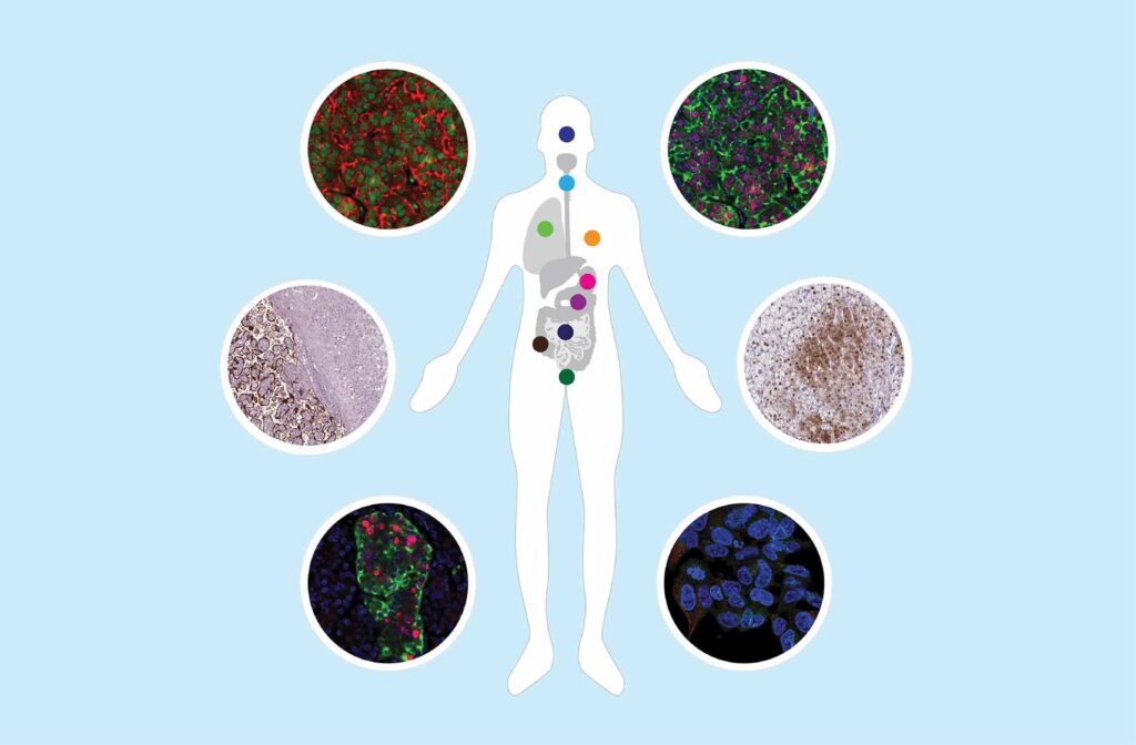
Understanding the development and progression of lineage cell types within the nervous system is critical for understanding the mechanisms of neurodegeneration and other brain diseases and can help identify potential sites for intervention. PrecisA Monoclonals™ and Triple A Polyclonals™ are enhanced validated primary antibodies highly specific to their target developed by Atlas Antibodies.
Below is a selection of markers targeting the neural lineage development pathways such as:
>E-cadherin/CDH1
>C21orf33/HES1
>Nestin
>NOTCH1
>Occludin
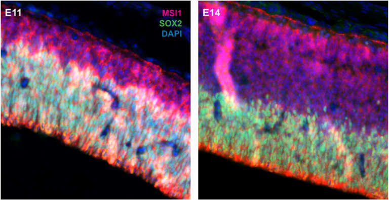
>SOX2
>FABP7/BLBP
>GFAP
>SLC1A3/GLAST
>C21orf33/HES1
>HES5
>PAX6
>SOX2
>Vimentin
Immature neuronal cells
>DCX
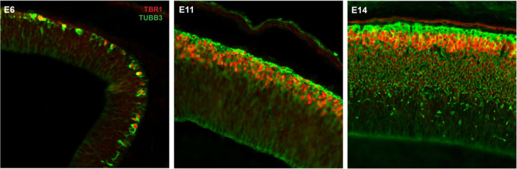
>TUBB3
>Nestin
>NeuroD1
>TBR1
Immature glial cells
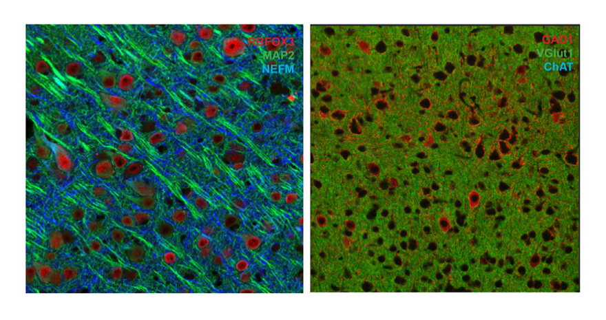
>RBFOX3/NeuN
>MAP2
>NEFM medium
>NEFM heavy
>synaptophysin
>PSD95/DLG4
Human Protein Atlas | Primary Antibodies | MolBoolean | PrEST Control Antigens | IHC | ICC | WB | ChIP | Enhanced Antibody Validation
We gladly support you by keeping you updated on our latest products and developments