Now you can you study cell behavior under confinement
10 October 2023
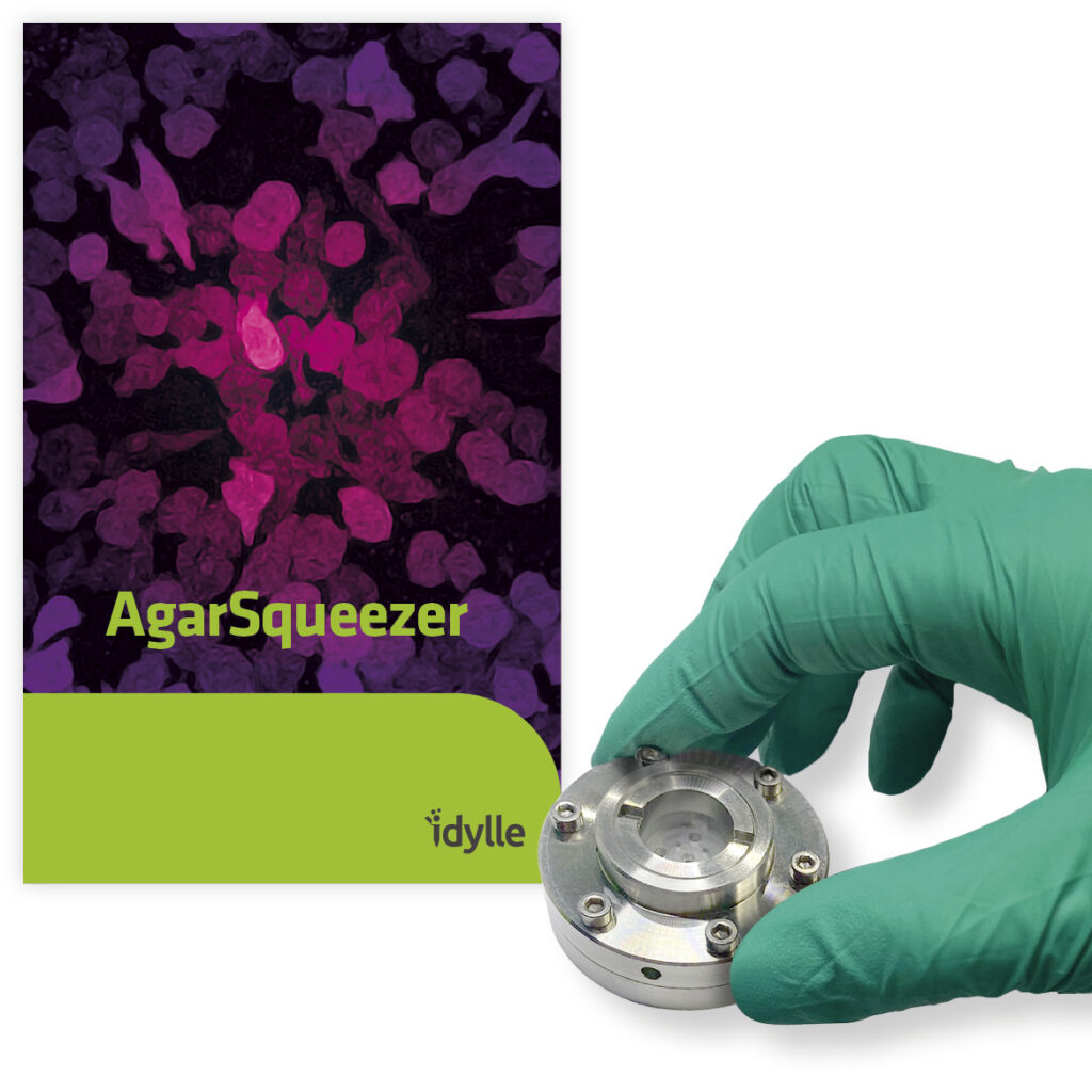
Just released by Idylle, AgarSqueezer helps confine your adherent and non-adherent cells and study their behavior within a physiological rigidity range.
AgarSqueezer is a microscope slide chamber equipped with a molded agar-based compression system. It is used to assess cell response to short and long-term mechanical confinement within a physiological rigidity range.
AgarSqueezer is very helpful if you want to analyze how your cells will react if you squeeze them for a prolonged period. Or if you want to study how mechanical confinement affects drug cell resistance. And if you want to perform immunostaining in situ.
Agarsqueezer has been successfully used with:
Human cells: primary T-lymphocytes, TF1 & ML2 leukemic cells, HS27A fibroblasts, MCF10A breast cells, MDA-MB-231 breast cancer cells, U-2 OS osteosarcoma cells, PC-3 & DU 145 prostate cancer cells, HT29 & HCT116 colorectal adenocarcinoma cells, HT1080 fibrosarcoma cells and megakaryocytes
Tunable stiffness in a physiological range [1-150] kPa: Use of agarose as a cheap and biocompatible polymer
Open access to the reservoir: Possibility to add drugs, and reagents. Easy medium renewal
Autoclavable & reusable systems
Compatibility with multiple microscopy techniques: Confocal, spinning, super-resolution. Open access for microscope objectives. Use of optical glass coverslip to make cells grow
Easy to recover coverslip with cells for subsequent molecular analysis: FACS, qPCR, Western-Blot, Immunofluorescence (possible in situ)
Long-term analysis of the cell adaptation to confinement: Up to several days, for time-lapse studies
Study of the specific impact of mechanical loads on the biology of cells: Gas permeability of the system allows to get rid of the hypoxia conditions
Easy to assemble and disassemble the system.
AgarSqueezer was developed by Audrey Prunet, Gilles Simon, Hélène Delanoë-Ayari, Véronique Maguer-Satta and Charlotte Rivière.
Audrey Prunet, Gilles Simon, Hélène Delanoë-Ayari, Véronique Maguer-Satta and Charlotte Rivière
Charlotte and Véronique, can you please tell us how and why you decided to transfer your invention?
Charlotte Rivière: “In the beginning, we wanted to analyze the influence of both stiffness and confinement on cells, mimicking highly confined situations such as fibrosis or cancer.”
Véronique Maguer-Satta: “On our side, our team was looking for a way to analyze the effect of long-term confinement with the ability to add a drug at any time. We all wanted to find a device that would also meet the requirements of subsequent classical molecular analysis (easy cell culturing and cells recovery, qPCR, western-blot, in situ immunostaining) as well as biophysical image-based analysis (high-resolution microscopy and video-microscopy). But we could not find any.”
Charlotte Rivière: “So all together, we decided to collaborate on the perfect set-up. We rapidly identified agarose as an interesting material to get medium and oxygen renewal, with no drug adsorption. We built several prototypes, improving precise control of the confinement, avoiding destruction of the gels and leakage of the culture medium! And then we came up with the Softconfiner device, that we published in Lab on a chip and decided to transfer with Idylle under the name of AgarSqueezer.
Today, we believe that the AgarSqueezer could be of interest for many researchers willing to better understand how mechanics can regulate cell behavior. We hope that researchers from different communities will also find it a useful tool!”
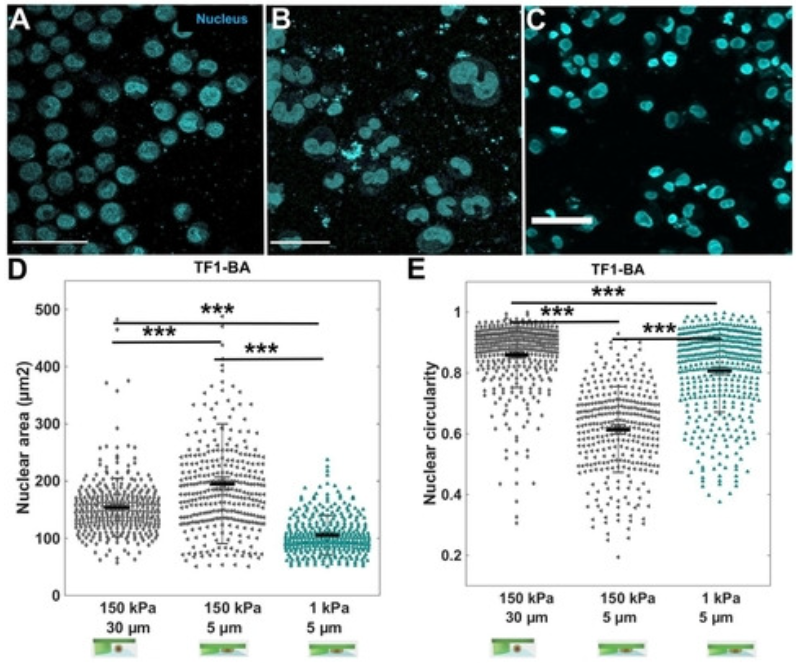
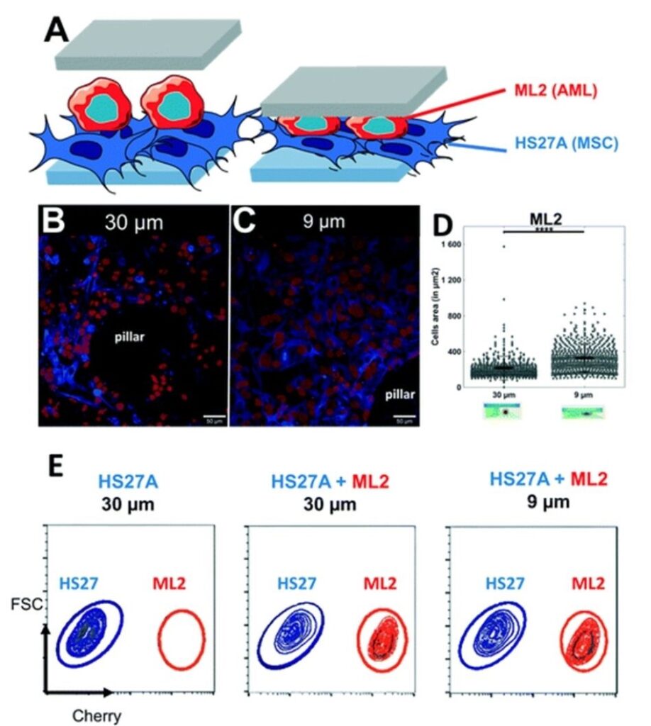
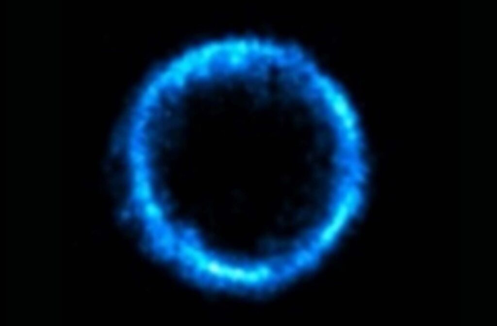
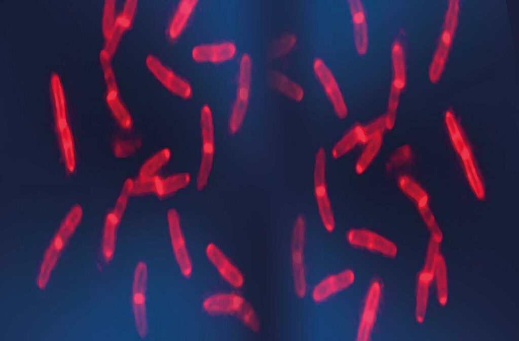
Influence of agarose stiffness on nuclear deformability. In situ immunostaining of TF1-BA cell nucleus (cyan) after 3 days in the soft cell confiner molded in (A) standard agarose (storage modulus of 150 kPa) with 30 μm pillars (no confinement); (B) standard agarose with 5 μm pillars (confinement) and (C) ultra-low agarose (storage modulus of 1 kPa) with 5 μm pillars. Scale bar = 100 μm. (D and E) Corresponding quantification of nuclear circularity and nuclear area of TF1-BA cells for the three conditions. At least 150 nuclei were analyzed per condition. Reproduced from Lab Chip, 2020,20, 4016-4030 with permission from the Royal Society of ChemistrySet-up mimicking the complexity of the tumor microenvironment. (A) Schematic representation of the two layers of cells inside the soft cell confiner. (B and C) Representative confocal images of leukemic cells (ML2, red) seeded on top of stroma cells (HS27A, blue) and inserted into the confiner with a height greater than both layers (h = 30 μm, unconfined, B) or smaller (h = 9 μm, confined, C) for 3 days. Scale bar = 50 μm. (D) Bar graph showing ML2 cell area quantification from unconfined (A, the average projected area of 216 ± 133 μm2) or confined (B, the average projected area of 329 ± 155 μm2). (E) Representative FACS plots following 3 days confinement in co-culture analyzed for cell content in HS27A-Turquoise and ML2-Cherry. Reproduced from Lab Chip, 2020,20, 4016-4030 with permission from the Royal Society of Chemistry
Co-Development, Production & Commercialization of Life Science Reagents & Technologies | Cell Culture | Microscopy | Bacteriology | Molecular Biology | Zebrafish | Exosomes