Uncover the structure of fibrinogen and its critical role in coagulation. Prolytix offers high-quality fibrinogen products, including research-grade reagents and antibodies, to support advanced scientific studies.
Plasma fibrinogen is a large glycoprotein (Mr=340,000) synthesized in the liver and circulating at a concentration of 2.6 mg/ml. It is a disulfide-linked dimer composed of three pairs of disulfide-linked non-identical polypeptide chains (Aα, Bβ, and γ).
Notable features of the Aα chain include the N-terminal peptide [fibrinopeptide A (FPA, 1-16)], factor XIIIa crosslinking sites, and two phosphorylation sites (generally 20-30% phosphorylation). The Bβ chain contains fibrinopeptide B (FPB, 1-14), an N-linked carbohydrate moiety, and an N-terminal pyroglutamic acid. The γ chain contains an N-linked glycosylation site and factor XIIIa crosslinking sites.
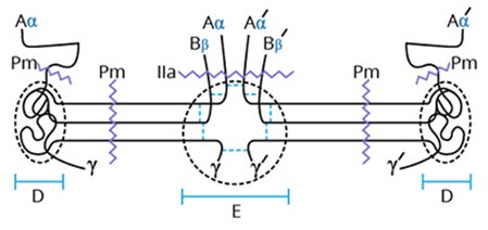
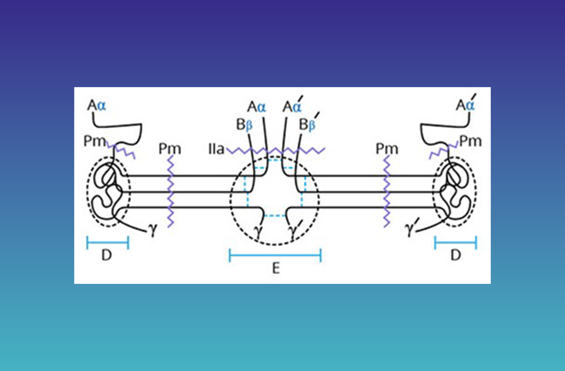
The two elongated subunits [(AαBβγ)2] are aligned in an antiparallel manner, forming a trinodular arrangement of the six chains. The nodes are formed by disulfide rings between the three parallel chains. The central node (n-disulfide knot, E domain) is formed by the N-termini of all six chains held together by 11 disulfide bonds. This region contains two thrombin-sensitive sites. The release of FPA by cleavage at R16-G17 generates fibrin I, exposing a polymerization site (17-20) on the Aα chain. These regions bind to complementary regions on the D domain of fibrin to form protofibrils. Subsequent thrombin cleavage of FPB (R14-G15) from the Bβ chain exposes additional polymerization sites and promotes lateral growth of the fibrin network.
Each of the two domains between the central node (E domain) and the C-terminal nodes (D domain) is composed of parallel α-helical regions of the Aα, Bβ, and γ chains coiled around each other to form a “coiled coil” with polar residues directed outward and nonpolar residues forming a hydrophobic core. In this region, all three chains possess a protease (plasmin) sensitive site. The other major plasmin-sensitive site is in the hydrophilic protuberance of the α-chain from the C-terminal node. Controlled plasmin degradation at these sites converts fibrinogen into fragment D and fragment E.
The thrombin-catalyzed cleavage of soluble fibrinogen to form fibrin is the terminal proteolytic event in the coagulation cascade. These soluble fibrin monomers spontaneously polymerize to form an insoluble fibrin network, which is stabilized by the factor XIIIa catalyzed crosslinking of Lys and Glu residues of α and γ chains. This fibrin network is the major protein component of the hemostatic plug. Fibrinolysis in vivo results in the release of two bound D fragments, D-dimer, from the fibrin mesh. D-dimer levels are used clinically as an indirect measure of fibrinolysis.
Prolytix has been providing customers with high-quality research reagents and custom collection devices for more than 35 years, including several products and services for fibrinogen and fibrin research that are discussed below and summarized in Table 1.
Table 1. Summary of fibrinogen and fibrin products available at Prolytix.
| Product Name | Prolytix Catalog # | Category |
|---|---|---|
| Human research grade fibrinogen | HCI-0150R | Human proteins |
| Human fibrinogen fragment D | HCI-0150D | Human proteins |
| Human fibrinogen fragment E | HCI-0150E | Human proteins |
| Human D-dimer | HDD-0151 | Human proteins |
| Mouse fibrinogen | MCI-5150 | Custom order, please inquire |
| Sheep anti-mouse fibrinogen | PAMFGN-S | Polyclonal antibodies |
| Sheep anti-porcine fibrinogen | PAPFGN-S | Polyclonal antibodies |
Contact one of our account managers to learn more about these reagents and others such as Prolytix custom collection devices in their extensive product catalog.
Coagulation & Hematology | Protein (human & animal) | Inhibitors | Fluorogenic Substrates | Antibodies | Factor Defcient Plasmas | Clotting Factors | Collection Tubes
We gladly support you by keeping you updated on our latest products and developments