Find products for your research
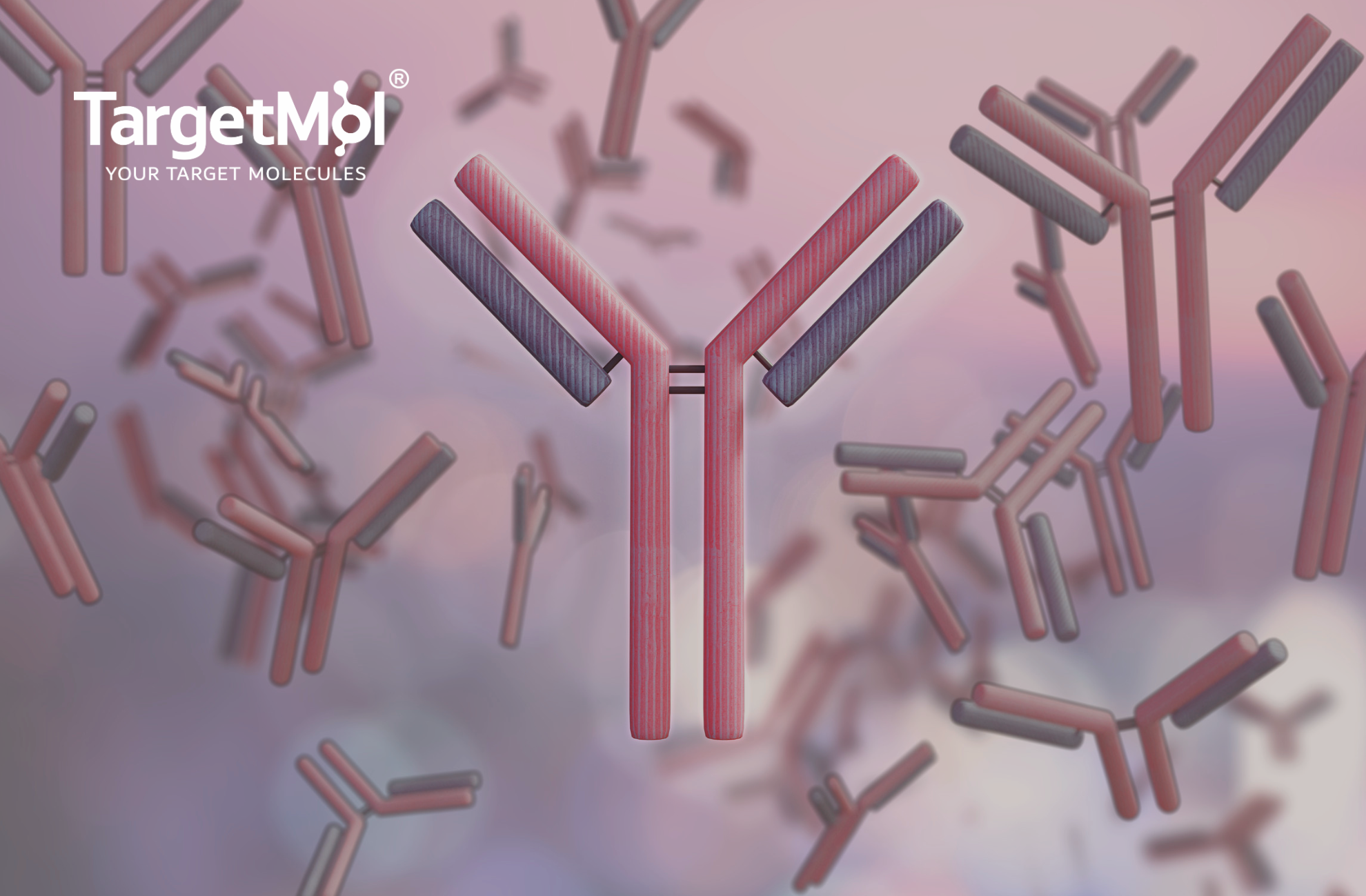
Immunology Research Solutions: Fuel Your Research with TargetMol
Discover TargetMol‘s comprehensive immunology research solutions, including inhibitors, agonists, and cytokines for innovative drug screening.
Discover how TargetMol’s advanced fluorescent dyes can provide you with precise, reliable, and intuitive visualization of cellular processes. Empowering every researcher, technician, and procurement specialist in the life sciences.
In cell-based experiments, fluorescent dyes have become powerful tools for revealing cellular structures and functions. Whether it’s changes in membrane potential, mitochondrial status, apoptosis, calcium signaling, or fluctuations in intracellular pH, fluorescence imaging technology provides us with precise and intuitive visualization methods. TargetMol currently offers over 2,600 dyes and dye-related products.
| Factors | Explanations |
| Emission Wavelength Compatibility | Compatible with microscope filter settings (e.g., FITC, TRITC, Cy5 channels) |
| Cell Permeability | Live-cell dyes should exhibit good cell permeability |
| Cytotoxicity | Non-toxic to cells, suitable for long-term observation |
| Targeting Specificity | Capable of specific staining of organelles such as the nucleus, mitochondria, endoplasmic reticulum, and lysosomes |
| Photostability | Resistant to photobleaching, enabling long-term imaging |
| Cell Membrane Staining | Some dyes, such as Hoechst, are suitable for fixed cells; others, like the Fluo series, are suitable for live cells |
| Catalog No. | Product Name | Target/Function |
| T6802 | CFSE | Cell Proliferation Tracking |
| T15458 | H2DCFDA | Intracellular ROS Detection |
| T14017 | 2-NBDG | Glucose Uptake |
| T2130 | Propidium Iodide | Dead Cell/Nuclear Staining |
| T6245 | BAPTA-AM | Calcium Chelator (Ca²⁺ Buffering) |
| T5840 | Hoechst 33342 | DNA Staining (Nucleus) |
| T15284 | FITC | General Labeling (Protein/Antibody) |
| TD0091 | Cy5.5 | Near-Infrared Dyes / In Vivo Imaging |
| T15129 | DiI | Cell Membrane Staining |
| T15609 | JC-1 | Mitochondrial membrane potential indicators |
| T2021 | Dihydroethidium | Superoxide anion (O₂⁻) detection |
| T18988 | FITC-Dextran (MW 10000) | Molecular weight labeling / permeability studies |
| T18977 | Dihydrorhodamine 123 | ROS detection (e.g., hydrogen peroxide) |
| T4139 | D-Luciferin potassium | Luciferase substrates (luminescence detection) |
| T36957 | BODIPY 493/503 | Neutral lipid (lipid droplet) staining |
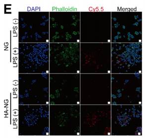
Li Y, et,al . Hyaluronic acid-coated polypeptide nanogel enhances specific distribution and therapy of tacrolimus in rheumatoid arthritis. J Nanobiotechnology. 2024 Sep 6;22(1):547. IF=10.6
RAW264.7 cells were divided into an LPS-treated group and an untreated group. NG and HA-NG were labeled with Cy5.5 (red). Cell nuclei were stained with DAPI (blue), and the cytoskeleton was stained with SF488-phalloidin (green). The final fluorescence staining results demonstrated the in vitro efficacy of HA-NG/TAC[2].
We gladly support you by keeping you updated on our latest products and the developments around our services.