Cloud-Clone offers a comprehensive product portfolio for exosome research, encompassing core products such as recombinant proteins, antibodies, and detection kits to meet the needs of the entire exosome research process.
For different research stages, Cloud-Clone recommends the following professional solutions:
Exosome Identification
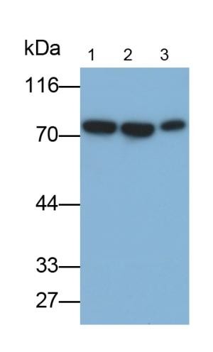
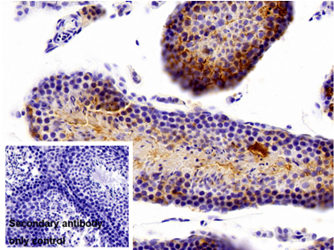
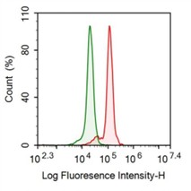
Utilize highly specific antibodies (e.g., anti-CD63/CD9 antibodies) in combination with ELISA kits for both qualitative and quantitative validation.
Mechanistic Research
Employ recombinant proteins (e.g., TSG101) along with functional antibodies to elucidate the biogenesis and functional mechanisms of exosomes.
Biomarker Screening
Multi-factor detection kits enable simultaneous analysis of multiple exosome biomarkers, significantly enhancing research efficiency.
Cell Microvesicles – Exosomes
Exosomes are membrane-secreted vesicular structures with a diameter of 30 to 150nm that are rich in specific protein, lipid, nucleic acid, and polysaccharide complexes. Exosomes are produced and released by almost all types of cells.
Exosomes contain a series of high-order oligomeric protein complexes that are closely related to cell membranes and exhibit significant molecular heterogeneity. These exosomes not only play an important role in protein quality control but also, upon release into the extracellular environment, exhibit multiple biological activities, such as reshaping the extracellular matrix and transmitting signals and molecules to other cells.
This intercellular vesicular transport pathway plays a crucial role in multiple aspects of human health and disease, including development, immune response, maintenance of tissue homeostasis, cancer progression, and neurodegenerative diseases. Based on the unique characteristics and functions of exosomes, they are currently being actively researched and developed as therapeutic agents for various disease models, indicating their broad application prospects in the future field of medicine.
Figure 1. Exosome System (The image is from Science).
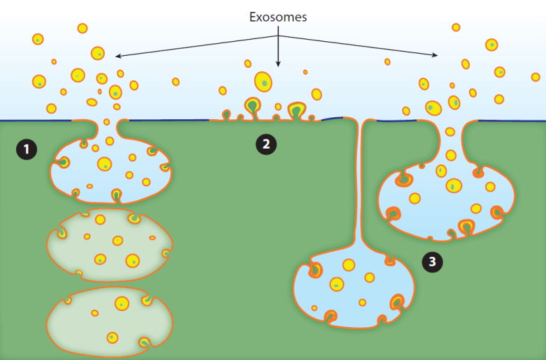
Biogenesis of Exosomes
Exosomes bud from endosomes and the plasma membrane. There are several modes of exosome biogenesis: 1. Inward invagination of the plasma membrane occurs, forming early sorting endosomes (ESEs) that encapsulate extracellular components and plasma membrane proteins. These ESEs can exchange materials with other organelles or fuse with each other to form late sorting endosomes (LSEs), which further develop into multivesicular bodies (MVBs) within the cell. MVBs release exosomes by fusing with the plasma membrane; 2. Exosome vesicles directly bud from the plasma membrane and are immediately released;3. Certain types of cells contain deep invaginations of the plasma membrane. These intracellular plasma membrane-connected compartments (IPMCs) are continuously connected to the extracellular environment through a neck region. The neck allows extracellular buffer and small molecular probes to pass freely but prevents the efflux of vesicles, making it a reservoir for vesicle accumulation. Subsequently, when the restriction at the neck of the IPMC is relieved, exosomes are released.
Exosome Inclusion Components
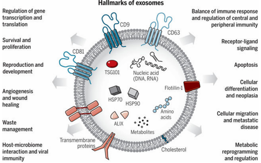
1. Exosomal Proteins
Exosomes contain a wide range of transmembrane proteins, lipid-anchored membrane proteins, peripheral membrane-associated proteins, and soluble proteins within the exosomal lumen. Among these, the tetraspanins (CD81, CD82, CD37, and CD63) are highly enriched in exosomes, whereas general plasma and lysosomal membrane proteins are not. Membrane proteins such as CD81, CD63, and CD9 are the most commonly used exosomal markers. Tetraspanins themselves lack catalytic activity but facilitate the transport, function, stability, and oligomerization of other membrane proteins. Many chaperone proteins associated with tetraspanins are present in exosomes, including major histocompatibility complex (MHC) class II proteins, immunoglobulin superfamily member 8 (IGSF8), intercellular adhesion molecule-1 (ICAM-1), etc., suggesting that these tetraspanins may mediate the functions of other proteins contained within exosomes. Exosomes represent a highly heterogeneous population with a unique ability to induce complex biological responses, with their heterogeneity primarily determined by exosomal proteins. For example, exosomes secreted by antigen-presenting cells contain high levels of MHC class II proteins and costimulatory proteins, whereas exosomes released by other cell types lack these proteins.
2. Exosomal Lipids
The outermost surface of exosomes is composed of a glycan corona attached to surface proteins and certain outer leaflet lipids. Beneath this glycan corona, the exosomal membrane contains phosphatidylcholine (PC), phosphatidylserine (PS), phosphatidylethanolamine (PE), phosphatidylinositol (PI), phosphatidic acid (PA), cholesterol, ceramide, sphingomyelin, sphingolipids, and some low-abundance lipids. The total lipid concentration of purified exosomes differs from that of plasma and other cellular membranes, and these differences may provide clues to the biogenesis mechanisms of exosomes.
3. Exosomal RNA
Exosomes contain RNA and are capable of transferring these extracellular RNAs (exRNAs) to other cells and tissues in a functional form. Relative to cellular RNA, exosomal RNA is enriched in specific RNA species, such as small non-coding RNAs (ncRNAs), including small nuclear RNAs (snRNAs), microRNAs (miRNAs), transfer RNAs (tRNAs), repetitive element RNAs, and fragmented RNAs. Specific RNA subsets bind to different targeted RNA-binding proteins to exert their functions.
4. Exosomal DNA
Exosomes contain DNA, including single-stranded DNA, double-stranded DNA, genomic DNA, mitochondrial DNA, and even retrotranscribed complementary DNA. The secretion of DNA via exosomes may contribute to DNA quality control in inflammatory regulation, potentially serving as a useful marker for cancer, viral infections, or chemotherapy resistance.
Roles and Research Prospects of Exosomes
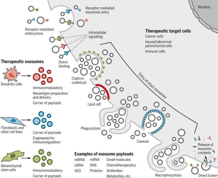
Exosomes serve as a means of intercellular material and signal communication. In disease research, exosomes represent an ideal drug delivery system with multiple advantages, such as low immunogenicity, good stability, and strong targeting capabilities. Exosomes lack the complexity of cells and organs, resulting in low immunogenicity. Unlike single proteins or small molecules, exosomes contain heterogeneous molecules, ensuring good biodistribution, pharmacokinetics, and cellular uptake of exosomes in the human body, making them easier to use for therapeutic purposes. As carriers of various therapeutic agents, exosomes can effectively protect the integrity of therapeutic drugs in vivo, greatly enhance drug bioavailability, reduce drug toxicity and side effects, and maximize the antitumor efficiency of drugs.
In summary, exosomes hold significant therapeutic and diagnostic potential in the medical field, and research into their properties and biological functions will unlock unlimited application prospects.
Figure 3. Cellular uptake of therapeutic exosomes. (The image is from Science).
Cloud-Clone has developed products related to the targets of this research. Some of these targets are listed as follows:

 Find products for your research