The developed brain organoid model, recently reported by researchers at Lund University in Nature Communications, enabled reproducible differentiation of functional neurons in 3D.
Advances in cell culture have made it possible to grow organoids, i.e. in vitro 3D tissues, aiming at resembling real tissue cell content and structure. Organoid differentiation protocols have been described for a variety of organs, including the digestive tract, kidney, and brain. However, the use of organoids can be limited by issues of reproducibility and incomplete cell maturation. Both these limitations were tackled by using Biosilk 3D matrix and a tissue specific Biolaminin in a recent publication by Dr. Alessandro Fiorenzano and coauthors at Lund University, Sweden [ref. 1].
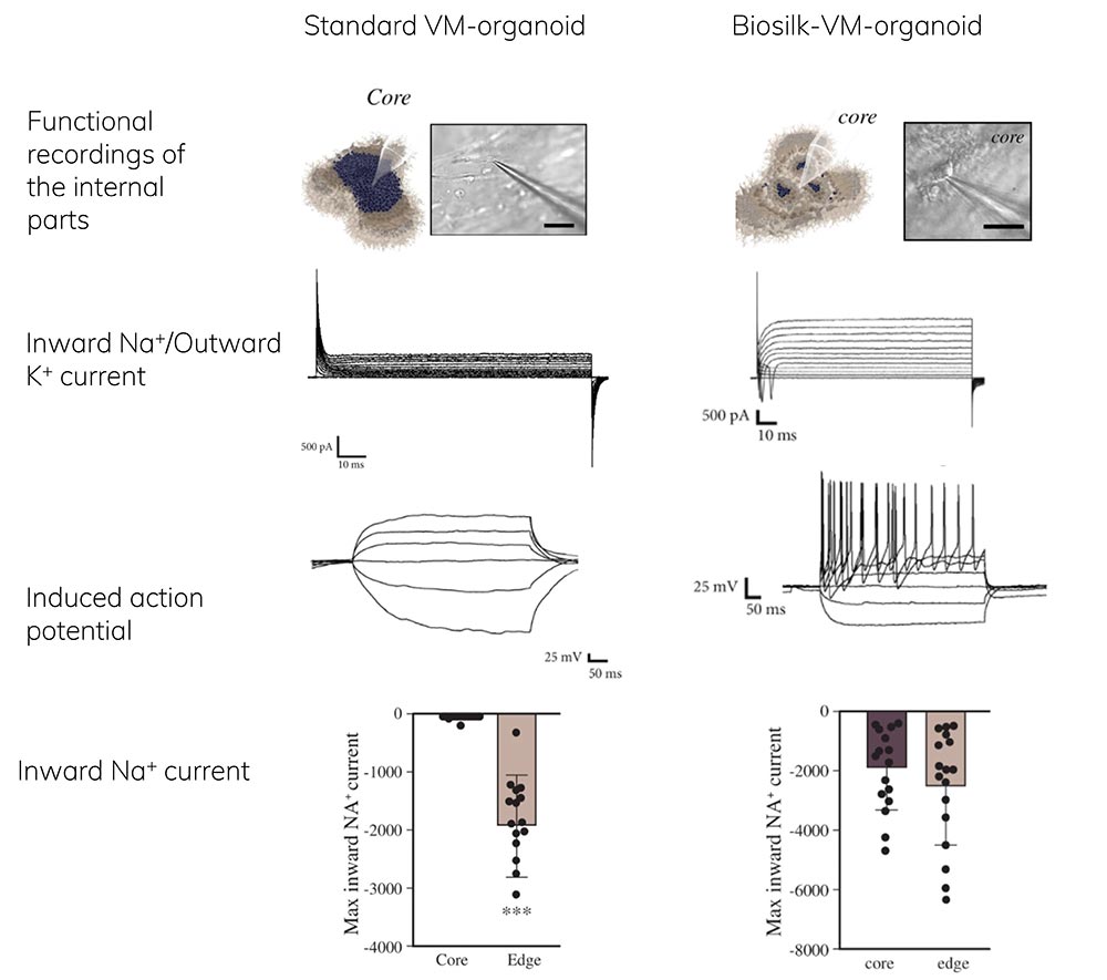
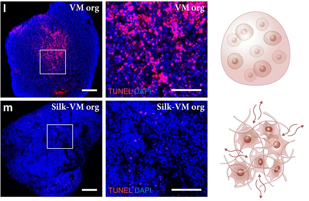
The authors characterized cell composition in the silk ventral midbrain (VM) 3D model and measured neuron cell activity, showing important improvements in experimental reproducibility compared to a widely used standard brain organoid protocol.
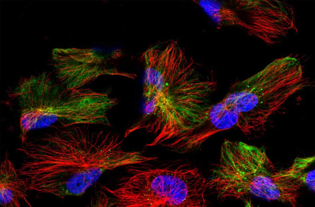
Their method resulted in efficient differentiation of pluripotent stem cells to functional midbrain neurons, homogenously distributed throughout the organoid. Remarkably, even cells inside the organoid were live and functional (Figure 1). This was achieved by using the Biosilk 3D matrix as the scaffold as it allows nutrients to flow through the tissue structure (Figure 2).
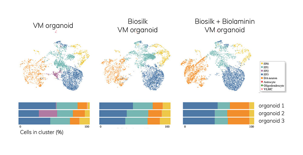
Single cell RNA sequencing and transcriptional profiling defined the cellular composition within the organoids, importantly revealing mature neurons and glial cells with high similarity to corresponding cell types in adult human midbrain. In addition, the authors showed increased similarity between the replicate organoids, improving the reproducibility of experiments (Figure 3). Adding a biorelevant Biolaminin substrate as silk coating significantly supported cell differentiation: for the reported VM brain application, Biolaminin 111 improved the number and transcriptional identity of dopaminergic neuron cells in the organoids.
The same research group has previously reported development of a GMP-adapted differentiation protocol for ventral midbrain dopaminergic progenitors for intracerebral transplantation, aiming for cure of Parkinson’s disease (ref. 2). Biolaminin 111 was used as the cell culture matrix to produce over 40 times higher yield compared to the original differentiation protocol.
Read more on the biorelevant laminin isoforms for neural cell culture.
Cell Culture Matrices | ECM | Biolaminin | Biosilk | Cell Therapy Grade Biolaminin 521 | 3D cell culture
We gladly support you by keeping you updated on our latest products and developments