Illuminate Metabolic Pathways with Luciferase Reporter Cells
The market for drugs treating metabolic dysfunction-associated steatohepatitis (MASH), obesity, diabetes, and other metabolic disorders is expected to grow dramatically, fueled by emerging therapies targeting the nuclear receptor thyroid hormone receptor beta (TRβ), or targeting G-protein Coupled Receptors (GPCRs) such as Glucagon-like Peptide-1 Receptor (GLP-1R).
To support drug development in these research areas, BPS Bioscience has developed a TRβ-GAL4 reporter cell line to interrogate TRβ signaling, and GPCR/CRE conditional reporter cells for drug screening and characterization. Indeed, cell-based assays are essential to the drug development process as they allow researchers to evaluate the biological activity of potential drug candidates, and to discover potential membrane permeability issues or interferences within a complex system that are not seen in biochemical assays.
Inducible reporter assays are an ideal option to study effectors of cell signaling pathways in the context of intact functioning cells. Luciferase reporters downstream of a pathway-specific promoter response element allow simple, robust, and quantitative measurement of signaling activity. They can be applied across a wide variety of cell types and signaling pathways and allow for high throughput screening of large libraries of compounds with inhibitory or activating function.
Applications
Identify new agonists or antagonists of a signaling pathway.
Screen compound libraries for molecules that selectively alter a target of interest or disease pathway.
Decipher the mechanism of action for a compound.
Study the parameters affecting a pathway of interest (analysis of function).
Advantages
Stable cell lines
Extensively validated
Provided with cell culture and validation protocols
Quantitative readouts
Suitable for high-throughput screening
GLP-1R, GIPR, and GCGR/CRE Luciferase Reporter Cells
GPCRs are a large family of proteins involved in many biological processes. They represent important therapeutic targets since approximately 50% of existing drugs target a GPCR. GLP-1R agonists, in particular, made a big splash in the field when they were approved for clinical use to combat obesity.
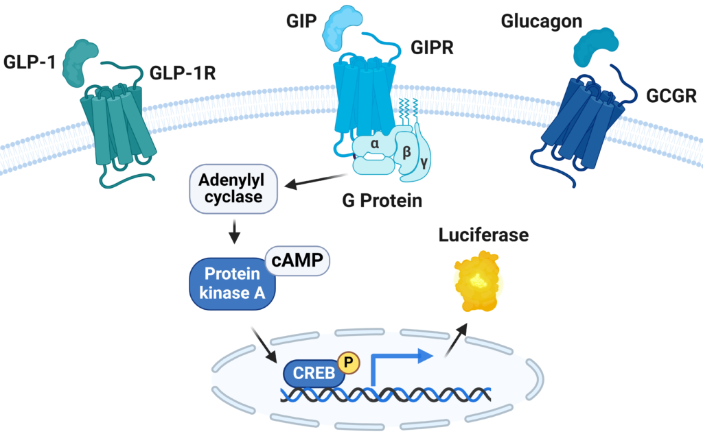
Figure 1: Illustration of the metabolism three cell lines
Three single clone, stable HEK293 cell lines were generated, each overexpressing a receptor and a Luciferase Reporter under the control of cAMP response elements responding to hormone and analog stimulation by increasing luciferase expression. Luciferase activity can be measured using a simple luminometer and is directly proportional to receptor activation. The three receptors are GLP-1R (78176), GIPR (Gastric inhibitory polypeptide receptor, 78589), or GCGR (glucagon receptor, 82187). Their three cell lines produce a robust luciferase readout upon stimulation of the cell surface receptor and are ideal for screening candidate molecules and determining their EC50.
Experimentally determined EC50 consistent with existing data
Strong induction signal: 40 to 300-fold stimulation depending on the cell line
Stable signal: luminescence maintained from 4 hours to 6 hours after analog addition
Amenable to high throughput: comparable results were obtained using 96-well and 384-well formats
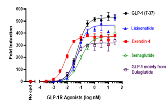
Figure 2: Dose-dependent response of various GLP-1 agonists in GLP-1R/CRE Luciferase Reporter cells.
TGFβ/Activin A/Myostatin-Responsive Luciferase Reporter HEK293 Cell Line
The activin A/myostatin pathway is of interest to scientists developing new therapeutics aimed at treating excessive weight, obesity, and diabetes, while mitigating the loss of muscle mass that comes with GLP-1 mimetics.
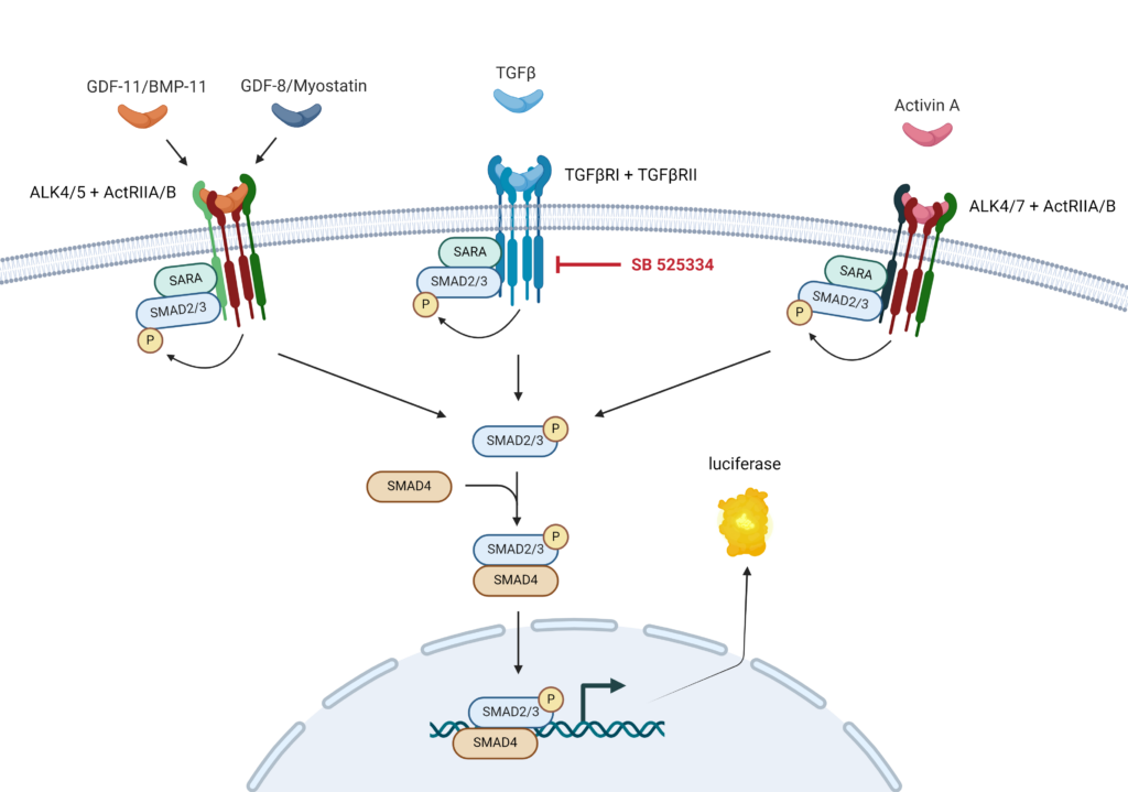
The TGFβ/Activin A/Myostatin-Responsive Luciferase Reporter HEK293 cells express the Firefly luciferase reporter under the control of SMAD-responsive elements (SMAD binding elements, SBE) and are ideal to monitor the activity of the TGFβ (transforming growth factor beta), Activin A, and myostatin-activated SMAD signaling pathway.
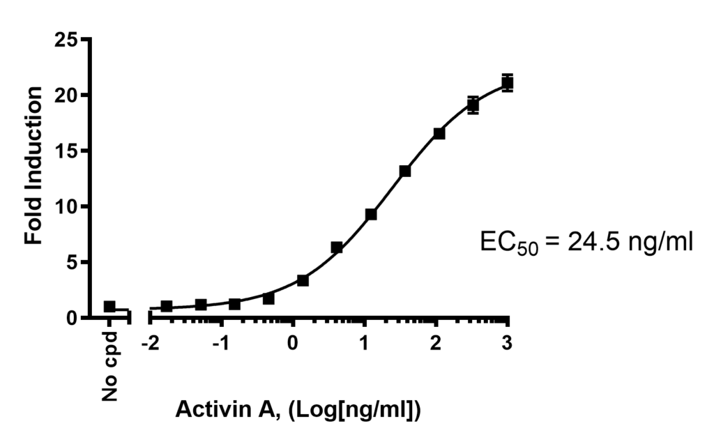
Validation experiments demonstrated that the cells respond to human TGFβ1 and to other cytokines of the TGFβ1 superfamily such as Activin A, GDF-8/Myostatin, and GDF-11/BMP-11. Luciferase activity induced by human TGFβ1 was decreased by SB525334, an inhibitor of the TGFβ1-receptor. Luciferase activity induced by Activin A, GDF-8/Myostatin, and GDF-11/BMP-11 was decreased by Bimagrumab, an antibody of the ActRII. Activin A induced luciferase activity was also decreased by Activin Blocker, an ActRIIA-Fc fusion protein, and an Anti-TGFβ antibody.
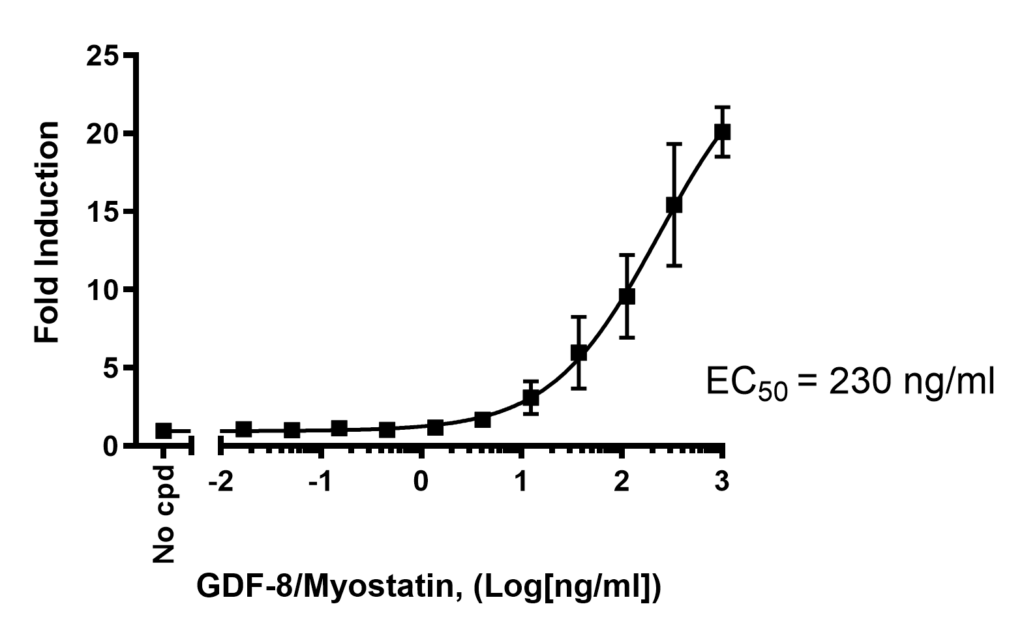
Dose-dependent increase in luciferase activity induced by myostatin (left) and activin A (right). Luciferase activity was quantified using ONE-Step™ Luciferase Assay System (60690).
The relative promiscuity of nuclear receptors makes it difficult to validate novel candidate drugs without interference from other nuclear receptors. The TRβ-GAL4 andTRα-GAL4 Luciferase Reporter HEK293 cells express Firefly luciferase under the control of the GAL4 upstream activation sequence (UAS), with constitutive expression of human thyroid receptor β ligand binding domain (TRβ-LBD) or the human TRα-LBD fused to the DNA binding domain (DBD) of GAL4 (GAL4-DBD). This system allows specific detection of thyroid hormone-induced activation of the relevant receptor with low cross-reactivity from other nuclear receptors.
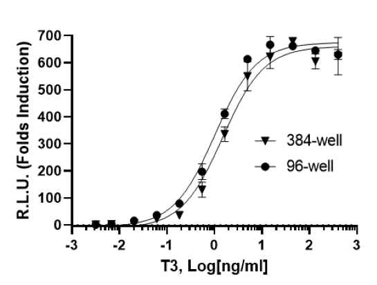
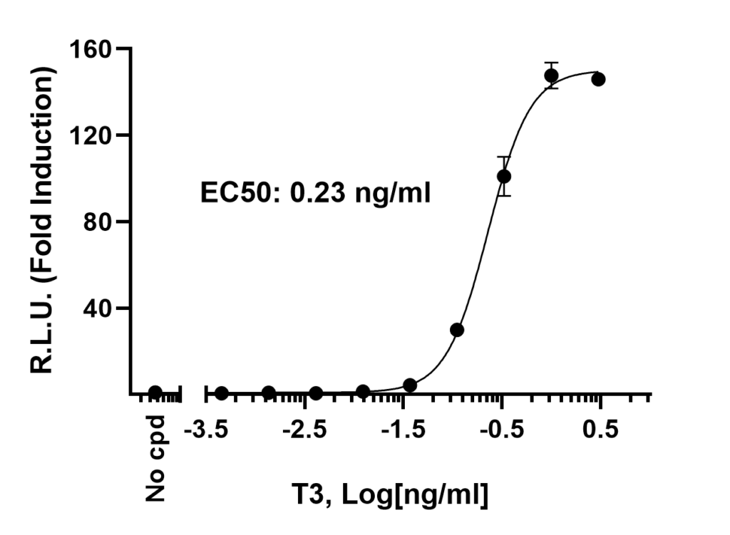
Both cell lines were shown to respond to stimulation with triiodothyronine (T-3).
Dose-dependent increase in luciferase activity induced by triiodothyronine (T-3) in TRβ-GAL4 (left) and TRα-GAL4 (right) Luciferase Reporter HEK293 Cell Lines. Luciferase activity was quantified using ONE-Step™ Luciferase Assay System (60690).
In addition to off-the-shelf cell lines, they offer rapid and dependable screening and profiling services for agonist screening or for determining an IC50. They also provide comprehensive cell line development services, should you not find your cell line of interest in our catalog. Contact us here.
Exceptional customer service
Project guidance and questions answered in a timely manner

 Find products for your research