MedChemExpress highlights how submerged, ALI and microfluidic co-culture systems help model the TME, immune evasion and drug responses.
In cancer research, co-culture systems have become essential tools for modeling interactions among multiple cell types within a controlled environment. Given the limited microenvironmental complexity of conventional tumor organoids, integrating immune and stromal components enables a more accurate recapitulation of tumor biology. Such systems enable the study of cellular crosstalk, signaling pathways, and tumor heterogeneity, and are therefore indispensable in modern cancer research[1].
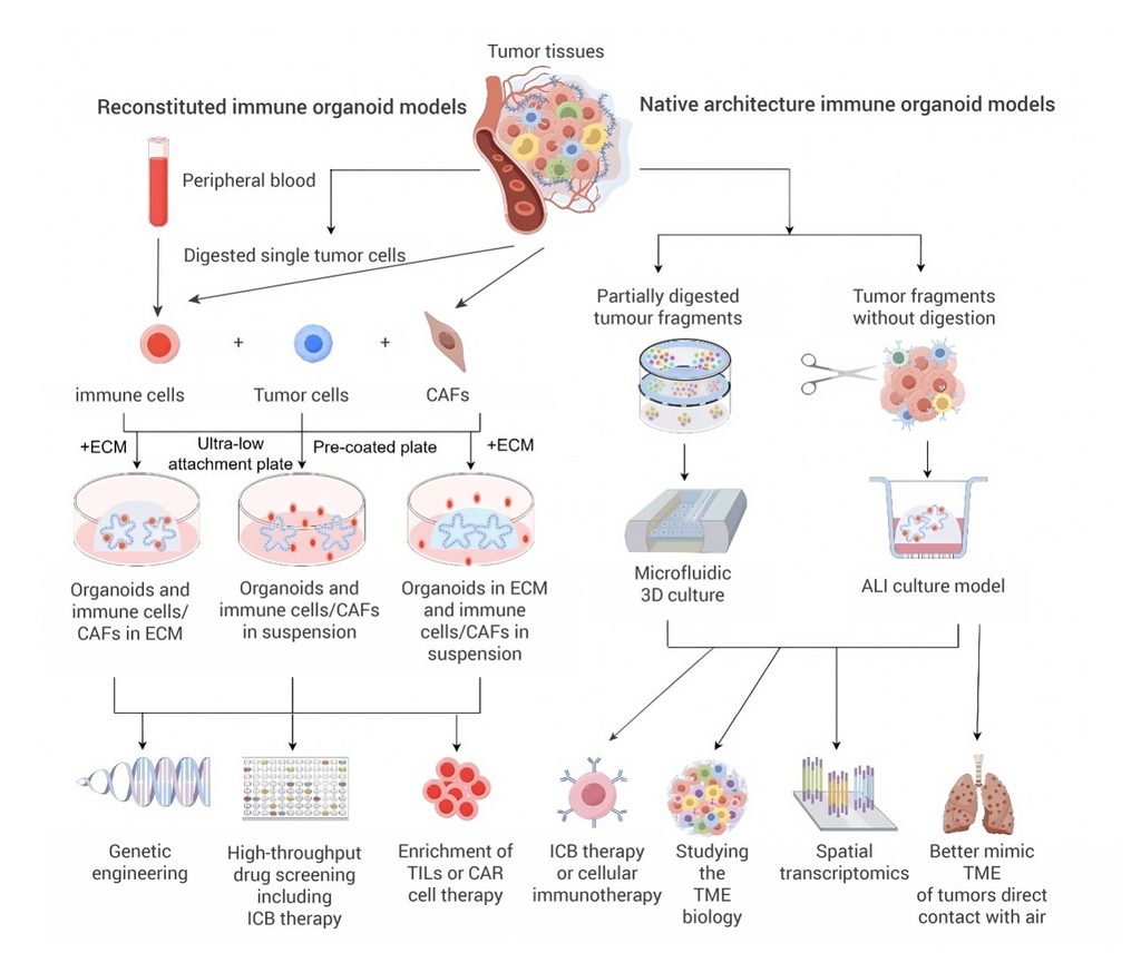
To capture cellular heterogeneity within the tumor microenvironment (TME) and heterotypic cell-cell interactions, three primary strategies have emerged: submerged immune cell-tumor organoid co-culture, 3D microfluidic culture, and ALI culture. Among these, submerged co-culture serves as a reconstructed TME model, while the latter two represent holistic TME models. The main characteristics and applications of different organoid models in simulating the TME are summarized in Figure 1.
Physical co-culture of immune cells with tumor organoids mimics immune cell infiltration in vivo, and is widely employed to investigate cytotoxic T cell activity and macrophage-mediated phagocytosis.
Matrix-embedded method: In this approach, immune cells and organoids are co-embedded within Matrigel or collagen matrices, preserving 3D architecture while allowing cell migration. Direct co-culture of pancreatic cancer organoids with T cells in Matrigel successfully induced the activation of cancer-associated fibroblasts.
Suspension co-culture method: Suspension co-culture using ultra-low attachment plates is suitable for high-throughput drug screening. For instance, co-culture of colorectal cancer organoids with CD8+ T cells facilitates rapid assessment of T cell cytotoxicity enhanced by PD-1 inhibitors.
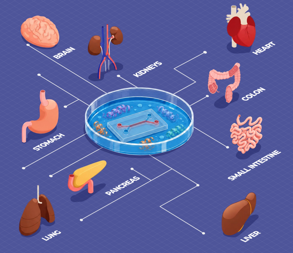
Microfluidic systems provide dynamic and biomimetic environments that enable precise control of biophysical parameters such as fluid shear stress and mechanical traction forces. In addition, these systems incorporate biochemical gradients to accurately emulate the physiological microenvironment of human organs. These systems overcome two fundamental limitations of conventional organoid models: the absence of physiological vascular perfusion and the lack of a functional immune system.
ALI culture is a 3D cultivation paradigm that simulates the luminal environment of hollow visceral organs. In this system, cells are seeded onto the permeable membrane of a collagen-coated insert; the basal surface remains submerged in the culture medium, while the apical surface is directly exposed to air, thereby mimicking in vivo epithelial architecture. Primarily applied to epidermal and respiratory epithelia, ALI culture allows airway epithelial cells to develop into pseudostratified layers with tight junctions, functional cilia, and mucin-producing capacities, faithfully recapitulating their native phenotypes. This approach has also been adapted to investigate interactions between organoids and TME.
Additionally, various co-culture models have been employed in cancer research, providing critical insights into the complexity of tumor behavior.
Table 1. Technological overview of co-culture models[2].
| Organoid type | Components used | Platform and matrix | Type of disease | Main outcomes | Detection technology | Reference |
|---|---|---|---|---|---|---|
| Cholangiocarcinoma organoid | T cell | Basement membrane extract-mediated direct co-culture | Cholangiocarcinoma | Allowing for the study of interactions between cholangiocarcinoma organoids and immune cells, as well as the evaluation of immunotherapies. | ATP quantification cell viability assay, HepG2 killing assay, Flow cytometry, Confocal time-lapse imaging and analysis, RNA sequencing analysis | [3] |
| Pancreatic ductal adenocarcinoma organoid | T cell | Matrigel-mediated direct co-culture | Pancreatic ductal adenocarcinoma | A resource for investigating pancreatic cancer and its microenvironment. | Flow cytometry, Immunofluorescence, Organoid histology, Immunohistochemistry, RT-PCR | [4] |
| Lung cancer organoid | T cell | Gel-liquid-interface co-culture model on a superhydrophobic microwell array chip | Lung cancer | Gel-liquid-interface co-culture can be used for developing diagnostic strategies for precision immunotherapies as well as understanding the underlying mechanisms. | Flow cytometry, qRT-PCR, Immunofluorescence, Drug sensitivity assays, Single-cell preparation and transcriptome library construction, RNA sequencing analysis | [5] |
| Clear cell renal cell carcinoma organoid | T cell | Microfluidic chip | Clear cell renal cell carcinoma | It offers insights into the rational design and optimization of viral-based immunotherapies for Clear cell renal cell carcinoma. | Oncolytic adenoviruses, Microfluidic chip assay, Immunofluorescence | [6] |
| Pancreatic ductal adenocarcinoma organoid | T cell | Matrigel-mediated direct co-culture | Pancreatic ductal adenocarcinoma | The combination of ultradeep single-cell sequencing and organoid techniques enabled rapid characterization of tumor-responsive T cell receptors for developing practical personalized T cell receptor-T therapy. | Immunohistochemistry, Immunofluorescence, RNA sequencing analysis | [7] |
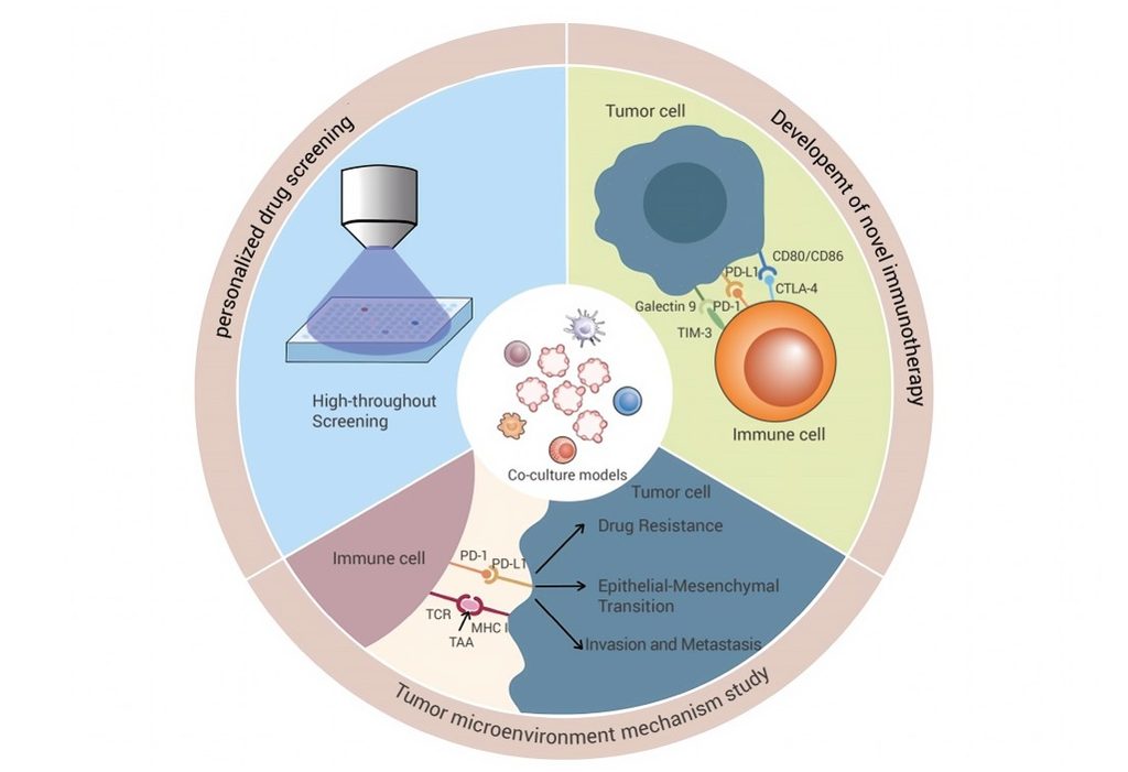
Tumor organoid-immune co-culture models have broad applications in oncology research and serve as indispensable experimental systems for investigating immune evasion mechanisms, elucidating the mechanisms of immunotherapy, and developing novel treatment strategies.
This figure illustrates the application scenarios of co-culturing tumor organoids with immune cells, including modeling interactions between tumor cells and immune cells, investigating TME mechanisms (such as immune responses and drug resistance), screening personalized therapeutics (e.g., high-throughput drug screening), and developing novel immunotherapies (e.g., immune checkpoint inhibitor development).
Ko et al. established 32 genetically engineered esophageal organoids and utilized single-cell RNA sequencing to profile their transcriptomic signatures and chemokine secretion patterns. Their study identified the concurrent deletion of TP53, CDKN2A, and NOTCH1 as a critical genetic determinant driving esophageal tumorigenesis and immune escape. These findings deepen our understanding of tumor immune evasion mechanisms and provide a theoretical foundation for the development of new immunotherapeutic approaches[8].
Furthermore, tumor organoid-immune cell co-culture models enable the efficient screening and evaluation of new immunotherapeutic drugs. Efficacy can be directly assessed by monitoring immune cells activation and subsequent tumor cell cytotoxicity in vitro. For example, Norkin et al. employed patient-derived colorectal cancer (CRC) organoids to screen several drug classes not traditionally used for CRC, including antipsychotic phenothiazines, cholesterol-lowering statins, antimycotic conazoles, selective estrogen receptor modulators, and antihistamines. They identified ifenprodil, opipramol, perphenazine, and toremifene as promising candidates with a favorable therapeutic window for targeting human CRC, warranting further in vivo validation[9]. Such findings provide significant preclinical data to support future clinical trials of these drugs.
Additionally, tumor organoid-immune cell co-culture models can be applied to personalized immunotherapy. By isolating tumor cells and autologous immune cells from a single patient, researchers can establish patient-specific co-cultures to empirically evaluate individual immune responses and the efficacy of tailored immunotherapeutic regimens. Van de Wetering et al. constructed patient-derived CRC organoids to test the activity of cetuximab in KRAS wild-type organoids. Their results, consistent with clinical observations, highlighted the potential of tumor organoids to directly assess the drug sensitivity of tumors in a personalized treatment approach[10].
Collectively, these research findings hold significant potential for informing personalized immunotherapy strategies for patients. The innovative integration of tumor organoids with immune cells offers a powerful tool to simulate the TME and interrogate complex tumor-immune interactions. This methodology not only deepens our understanding of tumor immune evasion but also establishes a vital experimental basis for discovering new immunotherapeutic strategies and personalized immunotherapy plans.
While tumor organoid-immune cell co-culture provides invaluable models for simulating the TME, their current methodologies heavily rely on matrices and culture media that present significant limitations:
Although current co-culture systems partially mimic the TME, achieving holistic physiological relevance remains challenging:
Despite existing challenges, tumor organoid-immune cell co-culture systems hold substantial potential:
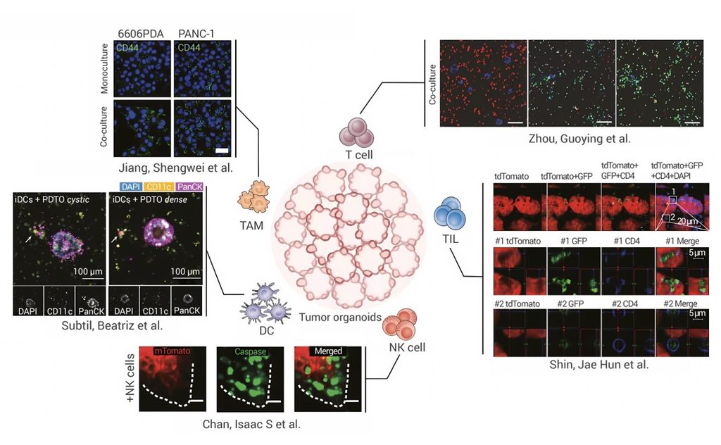
Figure 3. Future co-culture models[2].
Tumor organoid-immune cell co-culture systems substantially improve the biological fidelity of in vitro models by incorporating key cellular components of the TME. These models enable the investigation of tumor immune evasion mechanisms, support the screening of immunotherapeutic agents, and facilitate the development of personalized treatment strategies.
| Cat. No. | Types of tissue |
|---|---|
| HY-K6101 | Human breast cancer |
| HY-K6102 | Human Lung Adenocarcinoma |
| HY-K6103 | Human Small Cell Lung Cancer |
| HY-K6104 | Human Colorectal Cancer |
| HY-K6105 | Human Gastric Cancer |
| HY-K6106 | Human Cholangiocarcinoma |
| HY-K6107 | Human Cervical Cancer |
| HY-K6108 | Human Esophageal Cancer |
| HY-K6109 | Human Endometrial Cancer |
| HY-K6110 | Human Pancreatic Cancer |
| HY-K6121 | Human Head and Neck Squamous Cell Carcinoma |
| HY-K6122 | Human Bladder Cancer |
| HY-K6123 | Human Ovarian Cancer |
| HY-K6124 | Human Hepatocellular Carcinoma |
| HY-K6125 | Human Neuroendocrine Neoplasm |
| HY-K6126 | Human Kidney Cancer |
| HY-K6133 | Human Oral Squamous Carcinoma |
| HY-K6134 | Human Salivary Gland Cancer |
| HY-K6136 | Human Nasopharyngeal Carcinoma |
| HY-K6137 | Brain Glioma |
| HY-K6138 | Rhabdomyosarcoma |
| HY-K6139 | Neuroblastoma |
For researchers working with tumour organoids, immune co-culture systems or advanced 3D cell culture models, Bio-Connect can help you find the right MedChemExpress products for your work in the Benelux and Europe. Whether you are setting up a new model, comparing culture conditions or selecting reagents for drug screening, our team is happy to think along and point you towards suitable options.


Inhibitors | Agonists | Recombinant Proteins | Dyes | Assay Kits | Compound Libraries | Drug Screening | PCR | Cell Culture Reagents | Custom Synthesis, Screening, ADC, PROTAC Solutions
We gladly support you by keeping you updated on our latest products and developments