Bio-Connect supports organoid research with workflow solutions for starting cells, 3D culture, development, processing, engineering and analysis.
Get a clear overview of relevant product categories, trusted suppliers and portfolio highlights for every stage of the organoid workflow.
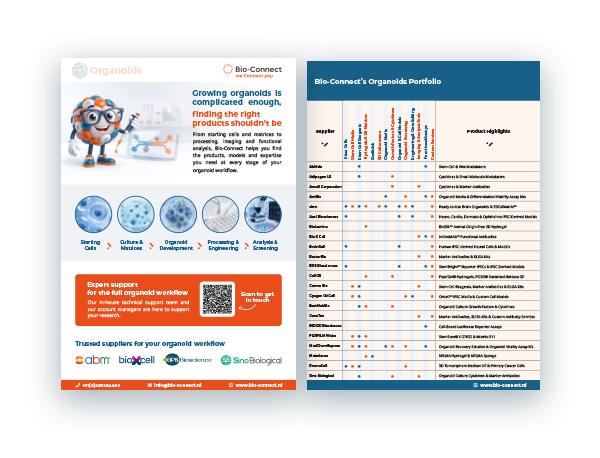
Use the workflow overview below to navigate through the key stages of organoid research. Click on a step to jump directly to the relevant section.
Organoid research depends on many connected workflow steps, including cell selection, 3D culture conditions, model development, processing and reliable analysis. Each step affects the quality, reproducibility and biological relevance of the final model.
Bio-Connect brings together products, models and services that support these workflow steps, helping researchers build more robust organoid models for disease modelling, drug discovery, functional screening and advanced 3D cell culture.
The type of starting cells, together with the signalling factors applied, strongly determines how an organoid develops and functions under specific culture conditions. Researchers currently rely on a few main cell sources, each with its own strengths and limitations.
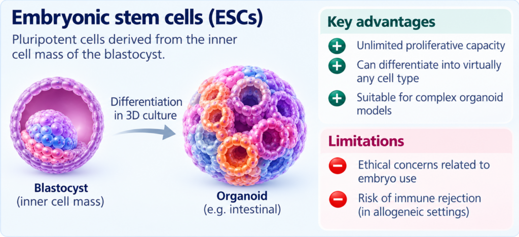
Embryonic stem cells are an important cell source in organoid research because they can proliferate extensively and differentiate into virtually any cell type in the body. This versatility makes them valuable for building complex organoid models. However, ethical concerns surrounding embryo use and the potential for immune rejection in translational applications can limit their use.
Induced pluripotent stem cells offer an alternative that can address some of these limitations. They can be generated from somatic cells and differentiated into derivatives of all three germ layers, making them suitable for a wide range of organoid types. When derived from individual patients, iPSCs can capture patient-specific genetic backgrounds and support disease modelling, drug discovery and personalised research. BPS Bioscience supports this field with engineered reporter iPSCs and iPSC-derived cell models that help researchers investigate disease mechanisms and evaluate therapeutic candidates.
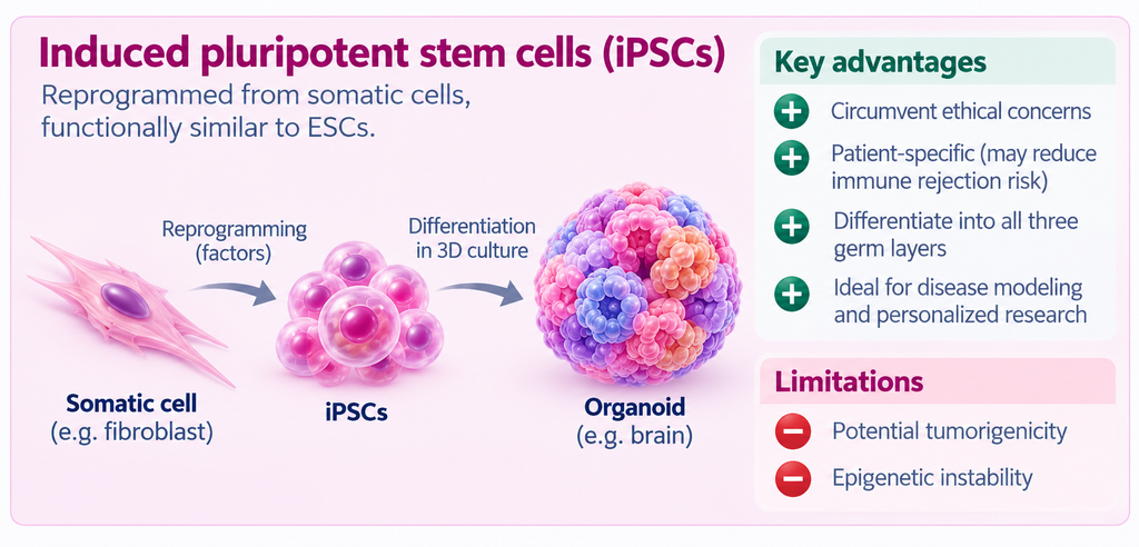
Cyagen complements this portfolio with custom iPSC generation from somatic cells, including blood-derived PBMCs and adult fibroblasts. These donor- or patient-specific iPSC lines can serve as starting material for disease modelling and directed differentiation into relevant cell types.
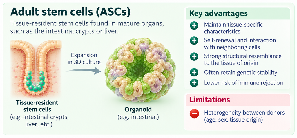
Adult stem cells follow a different principle. These cells reside within mature tissues, such as the intestinal crypts or the liver, where their self-renewal and differentiation are regulated by signals from the surrounding stem-cell niche. Under suitable culture conditions, ASCs can self-organise in interaction with neighbouring cells and generate organoids that retain important characteristics of their tissue of origin. ASCs are generally valued for their tissue-specific differentiation capacity and relative genetic stability, although their behaviour can vary depending on factors such as donor age, sex and tissue origin.
PromoCell provides well-characterised human primary cells and selected stem and progenitor cells. For selected cell types, donor information and HLA-typed material are available, helping researchers select appropriate cells for controlled and reproducible cell-based studies.
Finally, patient-derived cells can be isolated directly from patient material, such as biopsies or surgically resected tissue. These tissues can be processed using methods such as enzymatic digestion and fluorescence-activated cell sorting (FACS) to generate single-cell suspensions and enrich or isolate relevant cell populations, including cancer cells and, where present, cancer stem-like cells. This makes patient-derived cells particularly valuable for building patient-specific tumour organoids, which can preserve important genetic and histological features of the original tumour.
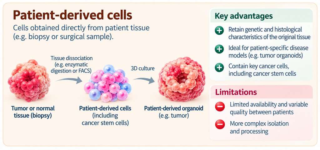
For researchers looking for flexible starting models, abm offers stem cell-derived cells, primary cells and engineered cell models, alongside custom cell-engineering services.
Once the right starting cells have been selected, the next challenge is providing an environment in which they can behave more like cells in native tissue. Traditional two-dimensional (2D) cell culture relies on nutrient-rich media to support cell growth, but it cannot fully reproduce the complex in vivo environment of tissues.
In three-dimensional (3D) culture, cells can form free-floating aggregates in suspension or grow within a supporting matrix, allowing them to interact and organise into complex structures. These conditions promote cell-cell interactions and signal transduction, enabling organoids to develop in ways that conventional 2D culture cannot support. Successful organoid formation depends on several interconnected factors, including cell selection, culture conditions, the microenvironment and cellular self-organisation.
At the centre of many 3D culture systems lies the extracellular matrix (ECM). A matrix does far more than simply hold cells in place. Physical properties such as stiffness and adhesiveness influence how cells self-assemble and organise, thereby affecting the morphology and function of the resulting organoid. Rather than acting as passive support, the matrix actively helps shape organoid development.
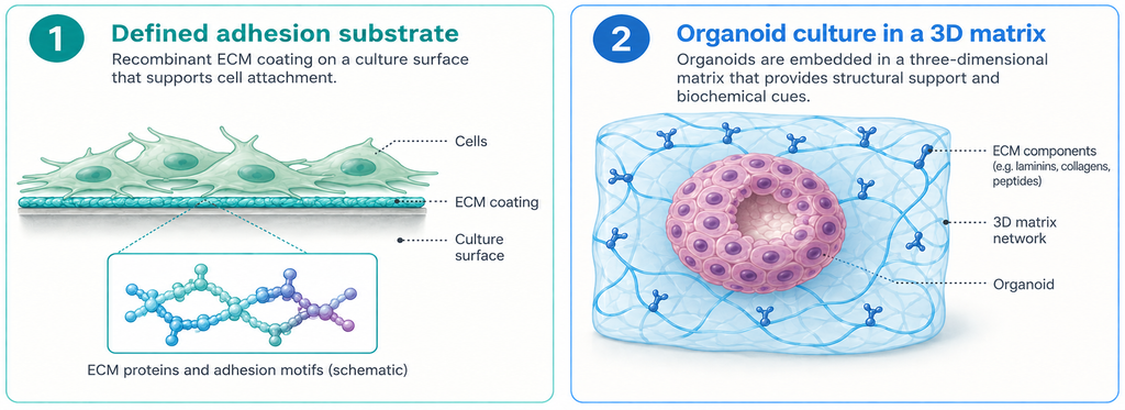
Importantly, there is no single matrix composition that is optimal for every organoid type, as requirements vary by cell source, model and protocol. In commonly used examples, intestinal organoids may be cultured in Matrigel, while some gastric organoid protocols combine Matrigel with collagen I, and some lung organoid protocols combine Matrigel with fibrin. By recreating selected physical and biochemical properties of the tissue microenvironment, these ECM components can help maintain epithelial polarity, support tissue-specific polarisation and promote processes such as airway branching.
Matrigel has long been used in organoid culture, but it also has notable limitations. It is derived from mouse tumour material, has an incompletely defined composition and can show batch-to-batch variability. Its cost can present an additional challenge. These limitations have encouraged the development of alternative matrix strategies, including recombinant and more defined ECM components, human-derived biomaterials and ready-to-use 3D culture matrices.
To support different matrix requirements, Bio-Connect offers access to a range of complementary solutions.
BioLamina provides full-length recombinant human laminins and defined 3D matrices for stem cell and organoid culture. For researchers seeking a human-derived, xeno-free approach, MetaTissue offers hydrogels and bioinks based on human proteins.
abm complements this portfolio with practical 3D matrix solutions such as 3DCelMatrix™, a ready-to-use extracellular matrix for organoid, spheroid and other 3D cell culture workflows.
For researchers requiring a defined cell-adhesion substrate, FUJIFILM Wako offers iMatrix-511, a recombinant laminin-511 E8 fragment used to support cell attachment and culture.
Kerafast provides additional cell culture tools, including 3D-printed cell culture inserts designed for controlled cell seeding, as well as media and reagents supporting hepatocyte culture and 3D culture applications.
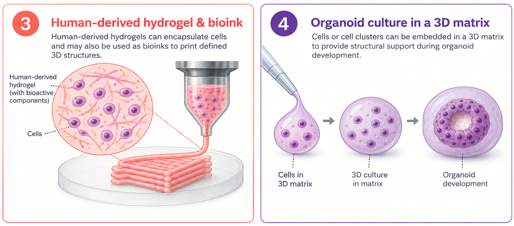
Together, these options allow researchers to move beyond a one-size-fits-all approach and select matrix properties, composition and origin according to the requirements of their organoid model.
Once cells are placed in a suitable 3D environment, their development into organoid structures depends on carefully controlled biological signalling. Growth factors, cytokines and small-molecule pathway modulators guide cells through self-organisation, proliferation, differentiation and maturation.
This process is highly coordinated. Cells integrate intracellular signalling with cues from neighbouring cells, the extracellular matrix and their mechanical environment, allowing them to migrate, communicate and organise into tissue-like structures. Growth factors, cytokines and small molecules are especially important because they activate, inhibit or modulate the pathways that influence cell fate and tissue organisation.
Each organoid type requires its own developmental recipe. In intestinal organoids, for example, Wnt3a, Noggin and EGF help maintain the Lgr5+ stem cell pool and support the formation of crypt-villus-like structures. This illustrates why organoid development depends on the right combination of pathway activation and inhibition.
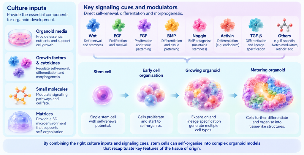
To support these signalling-driven workflows, Bio-Connect works with suppliers such as MedChemExpress (MCE), which offers small molecules, pathway modulators, organoid culture media and supplements; Sino Biological, which provides recombinant growth factors, cytokines and other proteins for cell stimulation and differentiation; and Cloud-Clone, which supplies organoid-related recombinant proteins, growth factors and cytokines, including key signalling targets such as EGF, BMP4, Noggin, RSPO1 and WNT3A.
Organoid maturation can take weeks to months, depending on the cell source and culture conditions. During this period, researchers evaluate whether the model develops relevant structural and functional features. Histology can reveal tissue-like architecture, functional assays can measure organ-specific outputs, and molecular methods such as transcriptomics and proteomics can show how closely the organoid resembles the original tissue.
As organoid models become more complex, researchers may also introduce additional cell types, co-culture systems or disease-specific conditions to better reflect the biological question being studied. These advanced models further increase the need for well-defined culture conditions and reproducible assessment methods.
After organoid formation, researchers often move into two complementary workflow stages: organoid processing and model engineering. Organoids may be recovered, dissociated, passaged, preserved or prepared for downstream analysis. Depending on the research question, cells or organoids may also be labelled or genetically modified to create reporter, knockout, knock-in or disease-specific models.
Although processing and engineering serve different purposes, both influence how reliably an organoid can be used in subsequent experiments. Careful processing can help preserve viability and model quality, while targeted engineering makes biological processes easier to track, manipulate and compare.
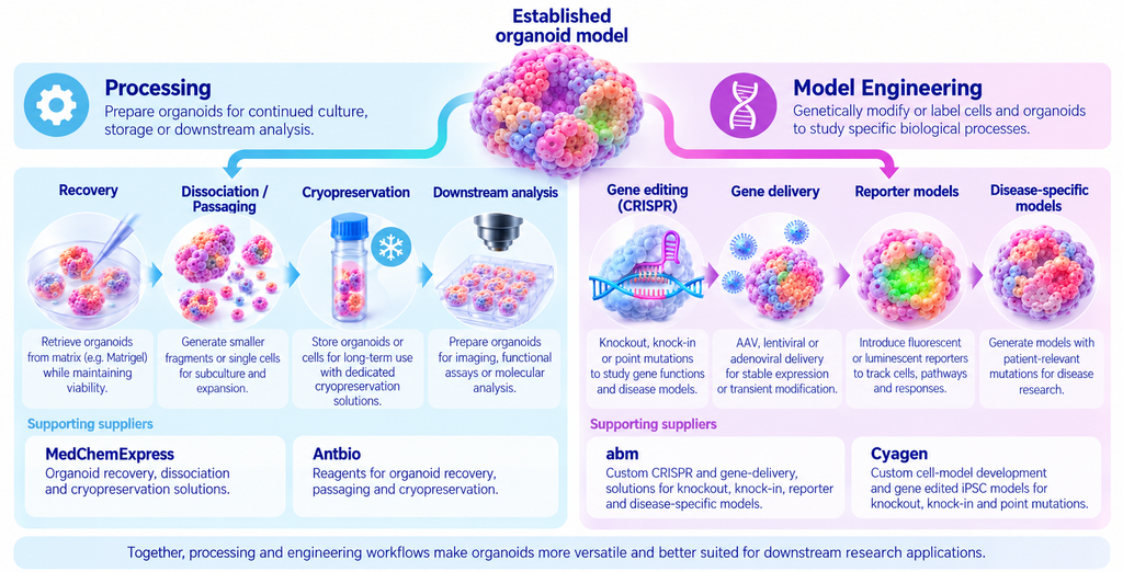
MedChemExpress offers reagents for organoid recovery, dissociation and cryopreservation. The Organoid Recovery Solution (HY-K6015) facilitates the recovery of intact organoids from Matrigel, while the Organoid Dissociation Solution (HY-K6013) enables passaging and the preparation of small cell clusters or single-cell suspensions. The serum-free Organoid Cryopreservation Medium (HY-K6014) is designed for the preservation of organoids and cell lines. Together, these products facilitate continued culture, storage and downstream analysis.
AntBio offers reagents for processing established organoid cultures, including collection, washing, recovery from hydrogels, passaging and cryopreservation. The Cell Recycling Solution (ABS90549) is described as an enzyme-free reagent that dissolves the surrounding hydrogel, enabling rapid recovery of organoids or cell spheroids for downstream analysis.
For model engineering, abm offers CRISPR and gene-delivery services, including knockout and knock-in cell line development, stable overexpression models and vector design. Available delivery options include AAV, lentiviral and adenoviral vectors, alongside gene-editing workflows with screening and validation. These solutions provide flexible routes for generating engineered cell models that can serve as starting points for reporter, disease-specific and other genetically modified organoid research.
Cyagen offers custom cell model development and specialised iPSC solutions, including gene-edited iPSC models for knockout, knock-in and point mutation studies.
Once organoids have been established and prepared for experimental use, imaging and functional assays can be used to investigate how they respond to drugs, toxins and disease-relevant stimuli. These read-outs turn complex 3D models into measurable data for disease research, drug discovery and precision medicine.
Imaging provides insight into organoid morphology, growth and treatment response. In melanoma organoids, for example, 3D photoacoustic imaging has been used to monitor structural changes and assess responses to cisplatin and temozolomide. Combined with marker-based analysis, imaging can also help researchers identify specific cell populations, examine protein expression and visualise tissue organisation.
GeneTex supports organoid characterisation with antibodies for applications such as immunohistochemistry, immunofluorescence, Western blotting, flow cytometry and ELISA.
Boster Bio complements this portfolio with a broad range of antibodies and ELISA kits for biomarker detection and protein analysis.
Functional assays provide insight into organoid function and responses to experimental interventions. Depending on the model and research question, researchers can assess viability, toxicity, neuronal activity, metabolic function, pathway activation and drug response.
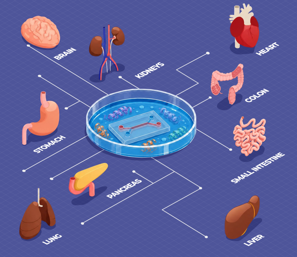
Cloud-Clone also supports organoid characterisation with antibodies, ELISA kits and multiplex assay kits for biomarker detection and target quantification. Its organoid-related portfolio includes markers for brain, liver, kidney, intestinal, lung, cardiac and tumour organoids, helping researchers assess differentiation, maturation, disease phenotypes and treatment responses across different model systems.
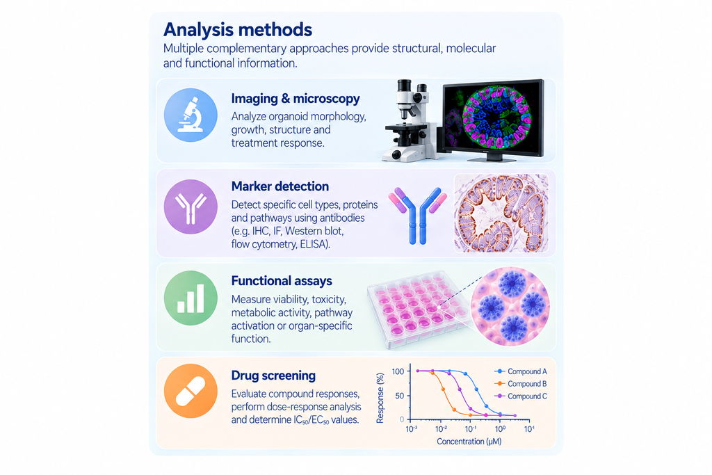
BPS Bioscience supports functional screening workflows with cell-based assays, reporter systems and screening services for investigating pathway activation and inhibition, receptor signalling and other functional responses. Its screening and profiling services include dose-response analysis and IC₅₀/EC₅₀ determination, helping researchers compare compounds, characterise initial hits and prioritise promising candidates.
For immune-related organoids and co-culture models, Bio X Cell provides functional antibodies for depleting specific immune cell populations and blocking, neutralising or activating biological targets. These reagents support studies of immune responses and mechanisms of action in organoid and organ-on-chip models.
Vector Laboratories complements these tools with lectins, lectin conjugates and glycan-detection reagents for glycobiology, immunohistochemistry, immunocytochemistry and immunofluorescence.
Jackson ImmunoResearch specialises in secondary antibodies and fluorescent antibody conjugates for protein detection and visualisation. Its reagents support applications including immunofluorescence, confocal microscopy, immunohistochemistry, immunocytochemistry and flow cytometry. Spectrally distinct fluorescent conjugates enable multi-colour labelling and the simultaneous visualisation of multiple targets within the same sample.
MyBioSource complements these capabilities with a broad selection of assay kits and reagents for assessing cell viability, cytotoxicity, proliferation, apoptosis and cellular metabolism. Its antibody portfolio supports biomarker detection and immunofluorescence-based characterisation, enabling researchers to combine functional cellular read-outs with molecular and phenotypic analysis.
More complex platforms can extend these applications further. Assembloids enable researchers to investigate interactions between different cell types or tissue regions, while organoid-on-chip systems introduce dynamic culture conditions and can support studies of drug efficacy and toxicity. Patient-derived models may also retain important genetic and disease-related features, creating opportunities to investigate patient-specific treatment responses.
A remaining challenge is the standardisation of analytical methods and functional read-outs. Reproducible imaging workflows, well-characterised reagents, robust assays and consistent evaluation criteria are essential for comparing results across experiments and laboratories.
By combining products for starting cells, culture systems, matrices, processing, model engineering and analysis, Bio-Connect helps researchers design more reliable and fit-for-purpose organoid workflows.
Get a clear overview of relevant product categories, trusted suppliers and portfolio highlights for every stage of the organoid workflow.
Wang, L., Chen, A., Zhang, D., & He, Z. (2025). From cells to organoids: approaches, regulatory mechanisms, applications, and challenges of organoids. Cells, 14(23), 1898. https://doi.org/10.3390/cells14231898
We gladly support you by keeping you updated on our latest products and developments